People regularly contact us to get our recommendations on books and other resources that we think are valuable to further ultrasound knowledge.
Whatever the topic, we’ve got our favourites – you can’t beat a good old fashioned book. In this list of top ultrasound books there’s something for everyone – from musculoskeltal to obstetrics. Plus – if you have a suggestion of a resource you have found valuable in your pursuit of ultrasound knowledge please let us know – we’d be happy to include it.
And a big thanks to you as each purchase through this webpage helps support us through affiliate partnerships.

POCUS in Critical Care, Anesthesia and Emergency Medicine
Hardcover – 19 March 2024
by Noreddine Bouarroudj (Editor), Peňafrancia C. Cano (Editor), Shahridan bin Mohd Fathil (Editor), Habiba Hemamid (Editor)
This book describes Emergency Ultrasound (EFAST echo, Lung Ultrasound, Vascular Ultrasound and Transthoracic Echocardiograhy) and all ultrasound technics in Anesthesia practice, i.e. Gastric Ultrasound, Airway Ultrasound and Ultrasound guided vascular access.
The book is divided into 11 sections and each section is devoted to a well-defined topic that are further investigated in the dedicated chapters richly and fully illustrated with colored and detailed sonoanatomy drawings. The volume represents a very useful, practical and highly didactic tool.
It presents concise up-to-date and evidence-based knowledge in POCUS and explains the use and benefits of ultrasound for intensivists, anesthesiologists, emergency specialists and allied professionals, as well.
Paperback – Illustrated, 11 June 2019
by Nilam J Soni MD MS (Author), Robert Arntfield MD FRCPC (Author), Pierre Kory MD MPA (Author)
Compact, hand-carried ultrasound devices are revolutionizing how healthcare providers practice medicine in nearly every specialty. The 2nd Edition of this award-winning text features all-new chapters, a greatly expanded video library, and new review questions to keep you fully up to date with the latest technology and its applications.
The Point of Care Ultrasound Handbook
Paperback – 21 June 2017
by Jason Bowman MD (Author), Jason Boitnott RN (Author), Branden Miesemer FP-C (Author)
This book is meant to be a reference for both the new and experienced point of care sonographer; to be a pocket guide to carry with you during your shift. We have included our best tips, tricks and any additional information that we have found helpful along our own journey towards point of care ultrasound nirvana. Throughout this book you will find helpful measurements and an additional advanced section so that this book can grow along with you as your POCUS skills increase. This book should be helpful for any paramedic, nurse, medical student, resident or attending physician learning or currently practicing point of care ultrasound.

The POCUS Manifesto: Expanding the Limits of the Physical Exam with Point of Care Ultrasound (POCUS)
Paperback – 27 October 2021
by Larry Istrail MD (Author)
This ‘why POCUS’ manifesto is an absolute must-read for anyone who uses a stethoscope.
“… In this manifesto, Dr. Istrail makes a solid case for why this technology should reboot the traditional heart and lung physical exam.” – Eric Topol MD, author Deep Medicine
‘Why do we continue to guess what diseases reside within the chest when we can look inside and know for sure?’ asks internal medicine physician Dr. Larry Istrail in this poignant and persuasive manifesto.
More than two centuries have passed since the 1816 inception of the stethoscope. During this time our medical care has advanced exponentially, while our physical exam has remained largely unchanged. That is until point-of-care ultrasound (POCUS) was brought to the patient’s bedside, enabling a clinician to digitally peel back the skin and observe the ecosystem of internal organs functioning in real-time. Through the lens of our physical exam origins, The POCUS Manifesto paints an inspiring landscape for the future of our physical exam and how POCUS plays a vital role in saving our patients from unnecessary complications, near-misses, and excess testing.
The POCUS Manifesto includes:
- Interviews with POCUS pioneers including Dr. Daniel Lichtenstein, the father of whole-body ultrasound, and Dr. Harvey Feigenbaum who discovered and developed modern echocardiography
- Insight from many modern-day POCUS experts including Dr. Renee Dversdal, Dr. Chris Fox, Dr. Nilam Soni, and many others
- A crash course in the history of the physical exam highlighting the work of Laennec, Auenbrugger, Hope, and many others
- A deep dive into lung ultrasound, how it was discovered, and what evidence exists to support its use
- A review of the literature on the accuracy of lung and heart auscultation
- An exploration of the evidence supporting the use of POCUS for diagnosing cardiopulmonary disease
- New modern-day techniques for evaluating the jugular venous pressure, tamponade, and many other cardiopulmonary diseases
Pocket Guide to POCUS: Point-of-Care Tips for Point-of-Care Ultrasound
Paperback – 15 February 2019
by Cameron Baston (Author), Christy Moore (Author), Elizabeth A. Krebs (Author), Anthony J. Dean (Author), Nova Panebianco (Author)
Pocket Guide to POCUS provides tips and reminders for issues and challenges encountered at the point of care. Questions such as what images to acquire, and how to get them are addressed as well as a quick glance at pathology in comparison to a normal image. For learners at all levels, this pocket guide will minimize the angst of scanning and prevent some of the more common pitfalls the authors have observed over their more than 50 cumulative years’ experience.

Cardiopulmonary Point of Care Ultrasound
Hardcover – 2 Oct. 2023
by Hatem Soliman Aboumarie (Editor), Marcelo Haertel Miglioranza (Editor), Luna Gargani (Editor), Giovanni Volpicelli (Editor)
This book is an illustration-rich practically focused resource on the use of cardiopulmonary point of care ultrasound (POCUS). It focuses on the applications of POCUS in a wide range of medical specialties especially critical care medicine, cardiology, emergency and respiratory medicine. Topics covered include the application of POCUS in diagnostics, monitoring of various cardiorespiratory conditions such as pulmonary edema, and the role it can perform in guiding interventions for pleural and pericardial effusion. Stunning illustrations and instructive accompanying video clips ensure that the reader can develop a deep understanding of how to properly apply the technology into day-to-day clinical practice.
Cardiopulmonary Point of Care Ultrasound is edited and authored by a group of world-renowned experts in the field of critical care echocardiography and POCUS from critical care medicine, cardiology, respiratory medicine, emergency medicine, surgery, radiology and cardiac physiology with a powerful multi-specialty and multi-disciplinary collaboration. It offers detailed insight into the applications of this technique and associated technologies across medical specialties with a focus on cardiorespiratory disorders. The user-focused emphasis in this resource makes it an ideal bedside manual for both experienced practitioners and trainees in these fields.

Basic Ultrasound Skills “Head to Toe” for General Intensivists (Lessons from the ICU)
Kindle Edition – 13 September 2023
by Chiara Robba (Editor), Antonio Messina (Editor), Adrian Wong (Editor), Antoine Vieillard-Baron (Editor)
The aim of this book, part of the European Society of Intensive Care Medicine (ESICM) textbook series, is to educate and train practitioners in the safe and competent use of diagnostic ultrasound imaging in the visualization and interpretation of different conditions. Specifically, the program will train practitioners in the safe and accurate acquisition of ultrasound images in the examination and monitoring of patients in intensive care settings.
The readers will be able to obtain knowledge on the use of ultrasound in a safe and effective manner and to learn how ultrasound examination can be used to optimize clinical management of patients.
The chapters include ultrasound physics, ultrasound examination technique, optimization of the image, bedside ultrasound diagnosis of different diseases. Different domains will be addressed from the evaluation of different organs such as the heart, the lung, the abdomen, vessels and brain, focusing on the basic skills required to an intensivists working in the general Intensive Care Unit settings.
The book – richly illustrated and with electronic supplementary contributions – is intended for anaesthetists and intensivists with basic knowledge of ultrasound physics and practical experience of ultrasound.
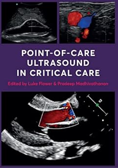
Point-of-Care Ultrasound in Critical Care
Kindle Edition – 1 August 2022
by Luke Flower (Editor), Pradeep Madhivathanan (Editor)
Point-of-care ultrasound has become a vital part of patient assessment in critical care units across the world. Its diagnostic sensitivity and specificity allow clinicians to accurately examine all major organ systems within a matter of minutes.
This book provides readers with a comprehensive, accurate and up-to-date practical guide to the use of POCUS in critical care, and features:
- detailed descriptions of how to perform a multi-system POCUS examination and its advantages over traditional techniques
- practical details of how to use POCUS in the assessment of shock, trauma and cardiac arrest
- contributions from leading POCUS specialists
- accreditation pathways
- hundreds of images
The book is accompanied by an additional image and video clip library which will help readers better understand ultrasound and incorporate it into their daily practice.
Point-of-Care Ultrasound in Critical Care is an essential handbook for clinicians working in critical care and anaesthesia. This book is also useful for those working in emergency medicine, internal medicine, surgery, and pre-hospital medicine. With POCUS likely to become a core competency in intensive care medicine over the coming years, this book is the perfect handbook for trainees.
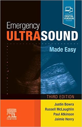
Emergency Ultrasound Made Easy
Paperback – 1 April 2021
by Justin Bowra MBBS FACEM CCPU (Editor), Russell McLaughlin MB BCH BAO FRCSI MMedSci FCEM CFEU (Editor), Paul Atkinson MB BCh BAO (Hons) MA FRCEM FRCPC CFEU (Editor), Jaimie L Henry MBBS BSc (Hons) MRCP (Editor)
This simple, jargon-free text fits in your pocket, providing an ‘on-the-spot’ guide to clinician-performed ultrasound in the emergency department, intensive care unit or in the field.
Written by an international team of experts and comprehensively updated in its third edition, Emergency Ultrasound Made Easy brings together in one volume the latest indications for focused ultrasound, including those related to the COVID-19 pandemic.
The text is highly accessible and easy to use in an emergency. It is aimed at the rapidly expanding cohort of non-radiologist clinical sonographers who use focused ultrasound. However, its broad scope (for example using ultrasound in the rapid diagnosis of DVT) makes it an invaluable addition to the library of any doctor with an interest in the technique, whether in primary care or the hospital setting.
- Simple to read and follow
- Free of jargon
- Fast step-by-step guide to ultrasound procedures
- Clear diagrams
- Tips and pitfalls to avoid
- Multiple accompanying videos featuring examples of ultrasound in clinical practice
- New chapter on the use of ultrasound in small anatomical structures such as the eyes and testes
- New chapter on paediatric ultrasound
- Respiratory chapter updated to include COVID-19
- Multiple accompanying videos featuring examples of ultrasound in clinical practice
- New chapter on the use of ultrasound in small anatomical structures such as the eyes and testes
- New chapter on paediatric ultrasound
- Respiratory chapter updated to include COVID-19
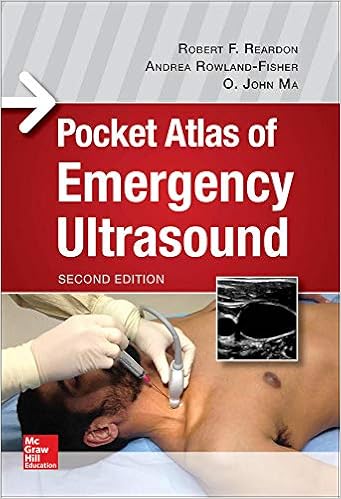
Pocket Atlas of Emergency Ultrasound, Second Edition
Paperback – Illustrated, 21 September 2017
by Robert F. Reardon (Author), O. John Ma (Author), Andrea Rowland-Fisher (Author)
The acclaimed quick-reference guide to performing and interpreting emergency ultrasound exams
More than 400 ultrasound images!
Spanning all body systems typically evaluated by ultrasound, this succinct, find-it-now pocket atlas allows you to instantly compare and contrast your real-time images with those appearing in the book. You will also find valuable how-to guidance on essentials such as probe placement, patient positioning, and proper settings – along with anatomical drawings that help you visualize affected organs. Featuring concise, bulleted text, and more than 400 ultrasound images and dozens of line drawings, Emergency Ultrasound, Second Edition is specifically designed to be used at the bedside in an emergency care setting.
Coverage includes:
- Clinical Considerations
- Clinical Indications
- Anatomic Considerations
- Technique and Normal Findings
- Tips to Improve Imaging Acquisition
- Common and Emergency Abnormalities
- Pitfalls

1st Edition, Kindle Edition
by John Christian Fox (Author/Editor)
There are already plenty of reference texts on how to perform a bedside ultrasound. Atlas of Emergency Ultrasound is different. It is a visually dynamic atlas, packed full of images of a broad spectrum of pathologic entities and emergency conditions. Over 300 detailed examples of positive ultrasound findings are provided, covering every organ system and showcasing the full range of pathology the clinician might encounter when using ultrasound. Each condition comprises several images with detailed captions and minimal text, enabling quick reference in a busy clinical setting. Both common and rare findings are included. A free companion website is also available (www.cambridge.org/9780521191685), featuring videos of cardiac, vascular and gastrointestinal ultrasound sequences and a range of ultrasound-guided procedures. Written by a leading emergency ultrasound physician and educator, and containing over 800 high-quality images, Atlas of Emergency Ultrasound is an invaluable resource for any clinician using bedside ultrasound.

Ma and Mateers Emergency Ultrasound, 4th edition
Hardcover – 22 October 2020
by O. John Ma (Author), James R. Mateer (Author), Robert F. Reardon (Author)
Ma and Mateer’s Emergency Ultrasound has been the definitive text for clinicians text since it was first published. This heavily illustrated guide from top experts in the field covers the training, techniques, and diagnostic skills for successful point-of-care ultrasound, with a special emphasis on problems most commonly encountered in the emergency or acute care setting.
Packed with 200 photos and illustrations, this brand-new edition gets you fully up to speed on the use of ultrasound in trauma, cardiac, critical care, pulmonary, hepatobiliary, renal, testicular, and other applications, as well as clinical considerations and indications, anatomical considerations, common abnormalities, and mistakes to avoid. With consistent chapter organization that makes it easy to find the answers you need, this peerless text includes side-by-side comparisons of normal/abnormal scans; new chapters on pulmonary and critical care; expanded chapters on cardiac and musculoskeletal ultrasound; and includes videos of common procedures in real time.
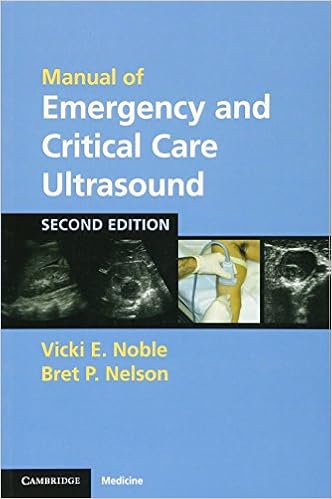
Manual of Emergency and Critical Care Ultrasound
Paperback – Illustrated, 16 June 2011
by Vicki E. Noble (Author), Bret P. Nelson (Author)
Ultrasound has revolutionized a physician’s ability to make urgent and emergent diagnoses at the bedside, and has changed the management of many acute injuries and conditions. This is a practical, concise introduction to what is rapidly becoming an essential tool for all critical care physicians: bedside emergency ultrasound. The Manual covers the full spectrum of conditions diagnosed using ultrasound and gives practical guidance in how to use ultrasound for common invasive procedures. Major applications are introduced using focused diagnostic questions and reviewing the image-acquisition skills needed to answer them. Images of positive and negative findings are presented, and scanning tips for improving image quality. The second edition has been substantially revised and expanded, with new images, updated literature reviews, new applications and clinical algorithms. New chapters cover additional procedures, musculoskeletal and pediatric applications, and the use of ultrasound in resuscitation. This text is invaluable for emergency physicians at all levels.
Emergency and Clinical: Ultrasound Board Review
Paperback – 14 April 2020
by Chiem (Editor), Dinh (Editor)
Emergency and Clinical Ultrasound Board Review is a comprehensive guide for preparing for the Advanced Emergency Medicine Ultrasonography or Critical Care Echocardiography board exams, and for residents preparing for in-training examinations in ultrasound.
The text consists of over 500 multiple-choice questions, organized into 18 chapters covering ultrasound topics such as physics, eFAST, echocardiography, thoracic, aorta, hepatobiliary, renal, pregnancy, soft tissue, ocular, procedural, airway, ENT, DVT, testicular, abdominal, and musculoskeletal applications.
This is a multi-specialty work, with contributors representing the fields of emergency medicine, internal medicine, cardiology, critical care, and radiology. Chapters include questions, answers with detailed explanations and references to primary or landmark articles to help better navigate a standardized exam. Questions are written in a case-based format that emulates the ABEM and NBE board exams, and are supplemented by over 800 figures, tables, boxes, and online videos.

Hardcover – 23 January 2014
by Philip Lumb (Author)
For physicians and nurses in critical care medicine, the increased demand and use of ultrasound necessitates further training. Critical Care Ultrasound helps meet that need. A straightforward, practical approach, an abundance of detailed ultrasound images and online video demonstrations provide step-by-step guidance on the principles and effective use of this important imaging modality in both diagnosis and assistance with specific procedures. Coverage includes the latest applications of ultrasound for neurologic critical care; vascular problems; chest; hemodynamic monitoring; and abdominal and emergency uses, as well as assistance in a variety of specific procedures in critical care medicine.
“…the book aims to and succeeds in fulfilling the appetite of different levels of expert in the use of ultrasound – from the beginner to the advanced practitioner.” Reviewed by British Journal of Anaesthesia, June 2015
“This book is a tremendous resource of practical knowledge and reference material. It will be of great help to trainees, critical care specialists, ICU nursing, allied health professionals, and anyone practicing acute medicine. Editors Philip Lumb and Dimitrios Karakitsos and the contributors are to be congratulated. “ Foreword by: Professor Teik E. Oh, AM , University of Western Australia, May 2015
- Incorporate a holistic approach. Visualize all or any parts of the body, tissues, organs and systems in their live, anatomically and functionally interconnected state and in the context of the whole patient’s clinical circumstances.
- See exactly how it’s done. Numerous ultrasound images and dozens of videos demonstrate the use of ultrasound in critical care.
- Rely on the guidance of more than 80 different experts from Australia, China, Middle East, Europe, USA, and Canada regarding the current and future use of CCU.
- Adapt the use of emergency ultrasound in specialized out-of-hospital (i.e., war zones, animals) and in-hospital (i.e., pediatric units) settings. Additionally, issues regarding CCU logistics, training, and education are analyzed for the first time.
- Access the complete contents, images, and video online at Expert Consult ―fully searchable!
Critical Care Ultrasound Manual
Paperback – 21 June 2013
by Anthony McLean B Sc(Hons) MB ChB MD FRACP FCICM FCSANZ (Author), Stephen Huang BSc MSc PhD CertEd DipLaw(LPAB) GDLP CRFS AMS(Cardiac) (Author)
Critical Care Ultrasound Manual is a concise step-by-step guide on the assessment of ultrasounds. It trains critical care physicians in applying Rapid Assessment by Cardiac Echo (RACE) and Focused Assessment with Sonography in Trauma (FAST) to sonography principles. Animated video clips of procedures assist the reader in comprehending the content covered in the manual.
- Focus on helping readers obtain rapid practical information to assist management decisions.
- User-friendly layout.
- Explanatory diagrams, ultrasound images enhance the learning experience.
- DVD showing video clips of procedures cross-referenced in the book.
- Practical tips and cautions are highlighted in Boxes.
- MCQs on each chapter allow readers to analyse what they’ve learnt.
- The Appendices provide a checklist to assist interpretation of transthoracic echocardiogram in a systematic way, and a chapter on Doppler principles for those who wish to prepare the way for Doppler measurements.
Ultrasound in the Critically Ill: A Practical Guide
Hardcover – 22 October 2021
by Andrew Walden (Editor), Andrew Campbell (Editor), Ashley Miller (Editor), Matthew Wise (Editor)
This book provides a practically applicable guide to the use of ultrasound in the care of acutely and critically ill patients. It is laid out in two sections. The first section attempts to take a comprehensive approach to specific systems of examination taking an organ focused approach covering techniques including Focussed Assessment with Sonography for Trauma (FAST) scanning and venous sonography. The second section presents a range of specific cases enabling the reader to develop an understanding of how to apply these methodologies effectively into their day-to-day clinical practice.
Ultrasound in the Critically Ill: A Practical Guide describes how to use ultrasound technologies in day-to-day clinical practice. Therefore, it is an ideal resource for all trainee and practicing physicians who utilize these technologies on a day-to-day basis.
Ultrasound for the Win!: Emergency Medicine Cases: 1
Paperback – 1 September 2016
by Dr. Jeffrey Shih MD (Author), Dr. Michelle Lin MD (Editor)
Ultrasound for the Win! Emergency Medicine Cases, Volume 1 is an educational series based on real Emergency Department cases where point-of-care ultrasound aided in the diagnosis or changed the management of a patient’s care! This collection of real jaw-dropping cases is geared towards anyone interested in learning more about point-of-care ultrasound! The cases included highlight several interesting and often surprising findings seen on point-of-care ultrasound that may have otherwise been missed! These exciting cases are expert peer-reviewed by Physician Leaders in the field of Point-of-Care Ultrasound including Dr. Chris Moore, Dr. Mike Mallin, Dr. Resa Lewiss, Dr. Mike Stone, and many more!
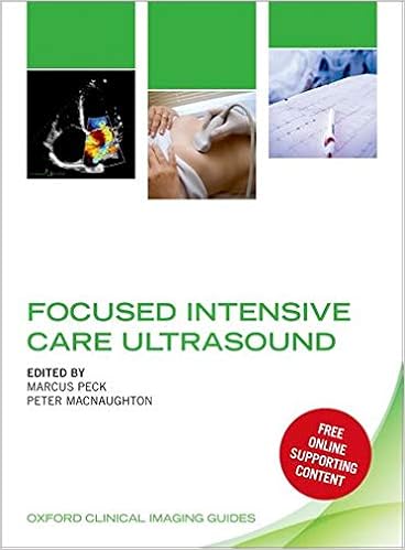
Focused Intensive Care: Ultrasound
Paperback – 28 March 2019
by Peck (Editor), MacNaughton (Editor)
A brand new series from Oxford University Press, the Oxford Clinical Imaging Guides are specifically designed to help doctors master imaging techniques. Each guide explains the principles and practice of using imaging in an easy-to-read, highly-illustrated, and authoritative manner. Focused Intensive Care Ultrasound is a practical manual designed to help you use ultrasound to assess and treat critically ill patients. Extensively illustrated, the book comes with online access to over 70 videos and 60 self-assessment questions.
This is the first book comprehensively mapped to the syllabi of Focused Intensive Care Echocardiography (FICE), Core Ultrasound for Intensive Care (CUSIC), Resuscitation Council UK’s Focused Echocardiography in Emergency Life support (FEEL), British Society of Echocardiography’s Level 1 Accreditation, and Focused Acute Medical Ultrasound (FAMUS). It is also ideal for European Focused Cardiac Ultrasound (FoCUS) training.
Providing a clear distinction between basic and advance skills, it introduces relevant aspects of advanced echocardiography for more experienced learners. With a focus on clinical issues, and supported by relevant physiological principles, this book teaches you exactly what you need to know to assimilate ultrasound into your everyday intensive care practice. Designed as an essential day-to-day reference and learning tool, this resource is ideal for trainees, consultants and critical care practitioners wishing to master ultrasound skills.
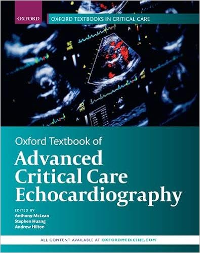
Oxford Textbook of Advanced Critical Care Echocardiography
Hardcover – 5 March 2020
by McLean (Editor), Huang (Editor), Hilton (Editor)
For critical care practitioners wishing to master advanced critical care echocardiography, it is necessary to master the theoretical aspects. The Oxford Textbook of Advanced Critical Care Echocardiography provides a physiological and evidence-based reference guide to the principles and techniques of advanced echocardiography.
Both transoesophageal and transthoracic echocardiography are addressed and over 300 videos, 470 figures and, 140 mcqs are included making this an ideal adjunct for courses for intensivists.The foundations of advanced echocardiography are clearly set out and specific assessment methods of critical care echo are outlined. As most critically ill patients do not suffer only one clinical problem a section on integrated techniques explains how to answer both common and unexpected critical care questions.
The Oxford Textbook of Advanced Critical Care Echocardiography includes access to online-only content, consisting of videos, figures and multiple choice questions that can be used to reinforce understanding. This online material can be accessed by activating your unique access code.
Authored by an international team of expert practitioners this textbook reflects the International Consensus Statement on Training Standards for Advanced Critical Care Echocardiography. Designed for trainees and consultants, this resource provides comprehensive and integrated coverage of all aspects of advanced critical care echo.
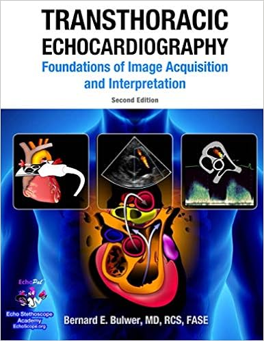
Transthoracic Echocardiography: Foundations of Image Acquisition and Interpretation. 2nd Edition
Paperback – 21 June 2018
by Bernard E Bulwer M.D. (Author)
This book is a the most highly illustrated and practical tour de force on the essential foundations of optimal image acquisition and interpretation in adult transthoracic echocardiography.
A foundation reference for cardiac sonographers, medical students, medical residents and fellows, specialists, and the medical imaging specialties.
FEATURES:
- The most highly illustrated guide to the ASE-recommended standards nomenclature in adult transthoracic echocardiography.
- An expert foundation guide to the performance and interpretation of the adult transthoracic echocardiography examination.
- Fundamentals of echo-anatomy, echo-physiology, hemodynamics, basic physical principles of cardiac ultrasound, and the standard echocardiographic views.
- Over 2,000 insightful illustrations, images, infographics, and flow charts.
WHAT’S NEW IN THE SECOND EDITION
- Greatly expanded Reference Guide, with updated Figures and Tables.
- Almost 100 new pages added with hundreds of new illustrations and images.
- Includes the most recent American Society of Echocardiography/European Association of Cardiovascular Imaging (ASE/EACVI) guidelines on chamber quantification, using highly illustrated formats.
- Future access to online updates and corresponding video clips.
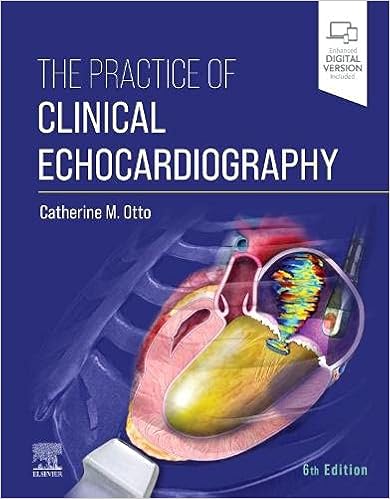
The Practice of Clinical Echocardiography
Hardcover – 27 August 2021
by Catherine M. Otto MD (Editor)
Ideally suited for those clinicians who have already mastered basic principles, The Practice of Clinical Echocardiography, 6th Edition, provides expert guidance on interpreting echocardiographic images and Doppler flow data. Through practical, clear, and carefully edited content, world-renowned expert Dr. Catherine M. Otto and her team of more than 65 leaders in echocardiography demonstrate how to apply advanced knowledge to daily clinical decision making. Newly reorganized sections cover advanced principles for the echocardiographer, best practices for echocardiography laboratories, transthoracic and transesophageal echocardiography, intraoperative and interventional echocardiography, and point-of-care cardiac ultrasound.
- Provides an in-depth, clear, and concise review of the latest clinical applications of echocardiography with an advanced level of discussion, now thoroughly updated with new clinical knowledge, new treatments and guidelines, the latest evidence, and innovations in advanced echocardiographic imaging.
- Reviews the technical aspects of data acquisition and analysis with an emphasis on outcomes.
- Covers key topics such as transcatheter interventions for valvular heart disease, prosthetic valve dysfunction, the athletic heart, cardiac assist devices, cardio-oncology, heart disease in pregnancy, advanced 3D echocardiography, strain imaging, stress echocardiography, and much more.
- Includes updated illustrations throughout―nearly 1,000 echocardiograms, Doppler tracings, anatomic drawings, and flow charts for diagnostic approaches―as well as hundreds of echo video clips keyed to images in the text.
- Discusses limitations, pitfalls, and alternate approaches.
- Features chapter summary boxes with new “Quick Reviews and a practical approach to echocardiographic data acquisition, measurement, and interpretation.
- Enhanced eBook version included with purchase. Your enhanced eBook allows you to access bonus images plus all of the text, figures, and references from the book on a variety of devices.
Textbook of Clinical Echocardiography
Hardcover – Import, 28 February 2018
by Catherine M. Otto MD (Author)
Today’s echocardiography continues to be a low-cost, minimal-risk procedure with the potential to yield a vast amount of detailed, precise anatomic and physiologic information. Dr. Catherine Otto’s Textbook of Clinical Echocardiography, 6th Edition, clearly outlines how to master the core principles of echocardiographic imaging in order to make an initial diagnosis and integrate this data in clinical decision making for patients with a wide range of cardiovascular diseases. Ideal for cardiology fellows, medicine residents, and cardiac sonography students, this bestselling text teaches all the essential elements of ultrasound physics, tomographic and 3D anatomy, image acquisition, advanced imaging modalities, and application in specific disease categories– all with a practical, problem-based approach.
- Concentrates on the foundational concepts you need to know to perform and interpret echocardiographic studies and to pass your board exams.
- Covers all advanced echo techniques, including contrast echo, 3D echo, and myocardial mechanics, as well as intraoperative and intra-procedural transesophageal echocardiography (TEE).
- Discusses what alternative diagnostic approaches to initiate when echocardiography does not provide a definitive answer.
- Includes new Echo Math boxes in each chapter for quick review and greater comprehension.
- Provides new ASE recommendations for chamber quantitation, including updated tables of normal measurements.
- Matches full-color anatomic drawings of heart structures with the 2D and 3D echocardiographic views.
- Pairs state-of-the-art echo images with more than 360 videos that illustrate the full range of cardiac disease diagnosed with this powerful imaging approach.
- Expert Consult™ eBook version included with purchase. This enhanced eBook experience allows you click on each figure to see the corresponding video, as well as search all of the text, figures, and references from the book on a variety of devices.
Textbook of Echocardiography for Intensivists and Emergency Physicians
Hardcover – 25 June 2019
by Armando Sarti (Editor), F. Luca Lorini (Editor)
The second edition of this superbly illustrated book equips practitioners with all the information that they will require in order to perform echocardiography appropriately and to interpret findings correctly. After clear exposition of fundamentals of the examination technique via both the transthoracic and the transesophageal approach, the functional anatomy of all normal and pathologic thoracic structures as observed on echocardiography is presented in detail.
A wide range of basic and advanced applications of echocardiography in the emergency, perioperative, and intensive care settings are then described. A final supplementary section considers further applications of ultrasound in the ICU, such as in patients with respiratory failure, trauma, or venous thrombosis.
This edition has been extensively revised and updated and includes entirely new chapters on various techniques and applications. It will be a tremendous aid to current best practice for all intensivists, emergency physicians, and anesthesiologists who intend to use echocardiography and ultrasound for diagnosis and guidance of optimal management in the critically ill.

Clinical Echocardiography Review
Paperback – Illustrated, 7 April 2017
by Klein (Author)
Perfect for exam review or clinical practice, Clinical Echocardiography Review: A Self-Assessment Tool, Second Edition features over 1100 self-assessment questions to keep you up to date with the latest advances and clinical applications in the field.
Written by national and international experts from the Cleveland Clinic and other leading institutions, this best-selling review tool offers a self-paced, highly effective way to assess and expand your knowledge of echocardiography and improve comprehension and retention of vital information.
Key Features:
- Over 1100 self-assessment questions with answers, many based on interpretation of sample images, make this the largest question-and-answer review in echocardiography.
- Over 340 online video clips enable readers to review real-life case studies to build knowledge for exam prep and clinical use.
- Fully updated throughout, with many new questions and answers, new images, and updated videos to keep you current.
- Complete coverage includes basics such as physics, artifacts, M-mode and Doppler echocardiography; clinically oriented topics such as atrial fibrillation, coronary artery disease, cardiomyopathies, and pericardial diseases; as well as dyssynchrony, 3D and strain evaluation and newer technologies such as LVAD and interventional echocardiography.
- Focus on congenital heart disease helps improve clinical diagnosis of these abnormalities.
- Special sections on valvular heart disease including prosthetic valves highlights the echocardiographic appearance of valve problems to speed diagnosis.
- Emphasis is placed on diagnostic interpretation throughout, benefitting cardiologists, fellows, anesthesiologists, and sonographers at every level of expertise.
Your book purchase includes a complimentary download of the enhanced eBook for iOS, Android, PC & Mac. Take advantage of these practical features that will improve your eBook experience:
- The ability to download the eBook on multiple devices at one time � providing a seamless reading experience online or offline
- Powerful search tools and smart navigation cross-links that allow you to search within this book, or across your entire library of VitalSource eBooks
- Multiple viewing options that enable you to scale images and text to any size without losing page clarity as well as responsive design
- The ability to highlight text and add notes with one click.
Paperback – 16 September 2016
by Sam Kaddoura BSc(Hons) BMBCh(Oxon) PhD DIC FRCP FESC FACC (Author)
Echocardiography (echo), the use of ultrasound to examine the heart, is a powerful and safe technique which is now widely available for cardiovascular investigation. This simple and highly praised text is a practical and clinically useful introduction to the subject. It aims to explain the echo techniques available, outlines what they are most suitable for, and most importantly puts echo into a clinical perspective. This book will be of value to all those who use or request echo, particularly doctors in training and medical students, but also physicians, surgeons, general practitioners, technicians, nurses and paramedics.
This Third Edition takes full account of recent advances in echocardiography. A new chapter on performing and reporting an echo has been added. New text has been added on the role of echo in individuals with cancer and in diseases of the aorta. There are updated and expanded sections on pregnancy, the continuity equation, diastolic function, long-axis function and 3-D echo. There are also updates and more detailed sections on the use of echo in emergency situations, in cardiomyopathies and pericardial diseases, in congenital abnormalities and in cardiac resynchronization therapy. Up-to-date published international guidelines have been referenced throughout.
New online content is available in the form of echo video images with accompanying self-assessment questions which will allow the reader to carry out self-assessment of knowledge and to see examples of the echos described in the text.
- This highly-praised book is a simple guide to a difficult subject, written in a conversational and accessible style. It is essential reading for anyone wishing to learn about echo – doctors in training, cardiac technicians, medical students etc.
- It provides full practical coverage of the clinical aspects of heart disease.
- It will be of great use to those experienced in echo both as a refresher and an accessible reference source.

Practical Approach to Transesophageal Echocardiography
Paperback – 24 July 2019
by Perrino & Reeves (Author)
With updated content and new expert contributors, A Practical Approach to Transesophageal Echocardiography, Fourth Edition is every clinician’s go-to reference for TEE skill-building. The book is written concisely, so you can quickly digest and apply critical information in a real clinical setting. Numerous figures throughout each chapter and hundreds of video clips on the eBook paint an important visual picture, illuminating the textual explanations and instructions. -Deals with topics and subjects for all levels of transesophageal echocardiography (TEE) usage and testing.
- Now with new information on interventional cardiology, transthoracic echocardiography, and the mitral valve.
- Brief self-assessments close each chapter, giving you the opportunity to test your skills knowledge immediately.
- More than 200 clinical and procedural video clips are available in the eBook bundled with this text. Enrich Your eBook Reading Experience -Read directly on your preferred device(s), such as computer, tablet, or smartphone.
- Easily convert to audiobook, powering your content with natural language text-to-speech.

Clinical Manual and Review of Transesophageal Echocardiography
Hardcover – Illustrated, 29 March 2019
by Joseph Mathew (Author), Madhav Swaminathan (Author), Chakib Ayoub (Author)
The acclaimed full-color clinical manual and review of TEE — completely updated to reflect the field’s latest breakthroughs and developments
Clinical Manual and Review of Transesophageal Echocardiography, Third Edition is written to be the field’s go-to resource and the standard reference textbook on the topic. It offers concise yet comprehensive coverage of the key principles, concepts, and developing practice of transesophageal echocardiography.
Completely updated, reorganized, and expanded, this Third Edition features a sectional format, each containing chapters that were reviewed and revised to provide a comprehensive discussion of physiology, pathophysiology, and echocardiographic approach for normal and common disease states. Where possible, important clinical information has been aligned with the principles of cardiovascular physiology and echocardiographic techniques. Narrative text, charts, videos, and graphs have been effectively integrated to provide rapid access to key clinical information for the purpose of improving clinical management.
Features:
- Addresses all the important clinical and technical issues and major exam topics
- Valuable appendices detailing normal chamber dimensions and volumes; wall motion and coronary perfusion; diastolic function; native valve areas, velocities, and gradients; and measurements and calculations
- Contributions from leading anesthesiologists, cardiologists, and cardiothoracic surgeons
- A complete practice exam and chapter-ending multiple choice questions make this the perfect review for board examinations in both basic and advanced perioperative echocardiography.

Ultrasound for the Generalist with Online Resource: A Guide to Point of Care Imaging
Paperback – Download: Adobe Reader, 31 December 2021
by Sarb Clare (Editor), Chris Duncan (Editor)
Point of care ultrasound is a critical tool required for assessing all patients, providing rapid answers to clinical questions and facilitating high quality care for patients. This essential guide caters for all generalist clinicians beginning their ultrasound journey and extends to more advanced assessments for those with established ultrasound experience wishing to advance their knowledge and skills. It covers a wide range of ultrasound topics from echocardiography, thoracic and COVID-19 to emerging areas such as palliative care, hospital at home and remote and austere medicine. An extensive collection of colour images, videos and examples of clinical applications will inspire readers to acquire the skills of point of care ultrasound quickly, safely and systematically. The printed code on the inside of the cover provides access to an online version on Cambridge Core. An essential aid for acute clinicians, paramedics, general practitioners as well as remote medical providers, medical educators and students.
Hardcover – Illustrated, 4 September 2020
by Bornemann (Author)
Make the most of point-of-care ultrasound in your primary care practice with this timely reference. The use of ultrasound in the primary care setting has grown dramatically in recent years, resulting in faster and more accurate assessment of patients, and safer in-office procedures such as joint injections and ultrasound-guided biopsies.
Ultrasound for Primary Care covers exactly what you need to know to incorporate or expand ultrasound in your practice – from when and how to use ultrasound to pearls and pitfalls to coding and billing issues.
- Timely coverage of this fast-growing field helps you effectively manage the whole patient and expand the scope of your practice.
- Case-based clinical vignettes put key concepts into the perspective of a busy practice, helping you determine when and how to best use ultrasound for optimal patient care.
- A consistent, templated format features chapters based on answering clinical questions commonly asked in the primary care setting.
- Ultrasound protocol includes probe placement illustrations, anatomic illustrations, and a complete description of the procedure, including an easy-to-follow algorithm as well as pearls and pitfalls.
- Billing codes and their descriptions are included for each procedure.
- Enrich Your eBook Reading Experience with Enhanced Video, Audio and Interactive Capabilities!
- Read directly on your preferred device(s), such as computer, tablet, or smartphone.
- Easily convert to audiobook, powering your content with natural language text-to-speech.
- Adapt for unique reading needs, supporting learning disabilities, visual/auditory impairments, second-language or literacy challenges, and more
Ultrasound Guided Musculoskeletal Injections
Hardcover – Illustrated, 18 September 2017
by Gina M Allen BM DCH MRCGP MRCP FRCR MFSEM DipESSR MScSEM (Author), David John Wilson MBBS BSc MFSEM FRCP FRCR (Author)
An ideal “how-to” guide for those who perform musculoskeletal injections, this unique multimedia resource by Drs. Gina M. Allen and David John Wilson demonstrates how to make the most out of the clear visualization provided by ultrasound-guided techniques. High-quality line drawings, clinical photographs, and ultrasound images clearly depict patient presentation, relevant anatomy, and sonoanatomy, and each technique is accompanied by a video showing exactly how to perform the procedure.
- Clear, concise text and numerous illustrations and videos make this reference your go-to source on today’s ultrasound-guided musculoskeletal injections.
- Bulleted text follows a quick-reference template throughout: clinical/ultrasound findings, equipment, anatomy, technique, aftercare, and comments.
- Each chapter covers the entire injection process with text on the left-hand page and corresponding images on the right.
- Every technique is supported with a video available online.
- Useful for clinicians in radiology, sports medicine, rheumatology, orthopaedics, pain medicine, and physical therapy – anyone who needs a clear, current guide to this minimally invasive treatment option for pain relief.
- Expert Consult™ eBook version included with purchase. This enhanced eBook experience allows you to search all of the text, figures, and references from the book on a variety of devices.
The Ultimate Guide to Point-of-Care Ultrasound-Guided Procedures
Hardcover – 12 December 2019
by Srikar Adhikari (Editor), Michael Blaivas (Editor)
This comprehensive book provides an in-depth examination of a broad range of procedures that benefit from ultrasound guidance in the point-of-care setting. It covers common procedures such as ultrasound-guided central and peripheral venous access to regional nerve blocks, temporary pacemaker placement, joint aspirations, percutaneous drainage, a variety of injections and airway management. Chapters examine a variety of topics critical to successful ultrasound procedures, including relevant sonoantomy, necessary equipment, proper preparation, potential complications, existing evidence and how to integrate these procedures into clinical practice. For each procedure, the book includes step-by-step instructions and discusses the advantages of ultrasound guidance over traditional techniques.
Providing rich procedural detail to help in clinical decision making, The Ultimate Guide to Point-of-Care Ultrasound-Guided Procedures is an indispensable, go-to reference for all health care providers who work in a variety of clinical settings including primary care, emergency department, urgent care, intensive care units, pediatrics, pre-hospital settings and those who practice in the growing number of new ultrasound programs in these specialties.

Ultrasound Guided Vascular Access: Practical Solutions to Bedside Clinical Challenges
Paperback – 15 December 2022
by Matthew D. Ostroff (Author), Mark W. Connolly (Author)
This casebook covers ultrasound guided vascular access with a focus on patient safety. Vascular access is the catalyst between provider and patient. Initially, patients were treated at hospitals for grave illnesses or terrible accidents; however, due to significant strides in medicine, the emerging hospitalized patient is now more commonly treated for chronic illnesses. For these patients, repeated hospital visits slowly deplete their vasculature, creating one of modern medicine’s greatest obstacles: vascular access. This book provides safe solutions to bedside clinical challenges from peripheral to tunneled central lines in the neonatal, pediatric, and adult patient populations.
Chapters present unique patient cases, incorporating the latest technology and techniques for safely and methodically meeting vascular access challenges, detailed discussions on therapeutic interventions, and trouble shooting. The book is structured so that each chapter builds on the last, offering peripheral, central, and tunneled catheter solutions through a standardized approach. Finally, each part features a COVID-19 case study including vascular access performed in the prone position.
This is an ideal guide for trainees, students, clinicians, and healthcare professionals performing ultrasound guided vascular access on patients.
Lung Ultrasound in the Critically Ill: The BLUE Protocol
by Daniel A. Lichtenstein (Author)
1st ed. 2016 Edition, Kindle Edition
Written by a pioneer in critical care ultrasound, this book discusses the basic technique and “signatures” of lung ultrasound and explains its main clinical applications.
The tools and clinical uses of the BLUE protocol, which allows diagnosis of most cases of acute respiratory failure, are first described in detail.
Careful attention is then devoted to protocols derived from the BLUE protocol – the FALLS protocol for diagnosis and management of acute circulatory failure, the Pink protocol for use in ARDS, and the SESAME protocol for use in cardiac arrest – and to the LUCI-FLR program, a means of answering clinical questions while reducing radiation exposure. Finally, the book discusses all the possible settings in which lung ultrasound can be used, discipline by discipline and condition by condition.
Lung Ultrasound in the Critically Ill comprehensively explains how ultrasound can become the stethoscope of modern medicine. It is a superb complement to the author’s previous book, Whole Body Ultrasonography in the Critically Ill.

The Pain Procedure Handbook: A Milestones Approach
Paperback – 31 December 2023
by Trent Emerick (Editor), Scott Brancolini (Editor), Michael E. Farrell II (Editor), Ajay Wasan (Editor)
This book fills the need for a succinct reference on how to master the progressive steps necessary to complete a wide range of complex pain procedures.
Organized by anatomic regions and target tissues, the book takes a structured approach to obtaining mastery of the steps required to perform a given procedure rather than providing more in depth or exhaustive reviews of theory and literature.
The book covers various nerve blocks and injections for treating chronic pain; how to correctly and safely perform the injection; feature x- ray or ultrasound pictures to help; and offer safety tips, and other pertinent information. Chapters begin with a brief summary, then define the specific skills necessary to perform a given procedure and deconstruct the procedure back into the individual skills expected by novice, intermediate, and advanced practitioners.
Practical and concise, The Pain Procedure Handbook is aimed for medical students, residents, fellows, and physicians interested in chronic pain medicine, anesthesiology, acute pain medicine, interventional radiology, and physical medicine and rehabilitation.

NYSORA Nerve Block Manual: First Edition
Paperback – 16 December 2022
by Admir Hadzic (Author)
NYSORA manual is a definitive guide to ultrasound-guided peripheral nerve blocks (PNBs) and interventional analgesia injections, written by Dr Hadzic and his top NYSORA team. It features complete and strictly practical information on the standardized, clinically most applicable techniques. The manual features only highly practical, richly illustrated information, instead burdening the reader with a literature discussions or non-practical considerations.
Here’s what you get in ONE source:
- Well-established, reproducible, ultrasound-guided techniques.
- Practical tips that are immediately applicable in clinical practice!
- Pragmatic instructions without burdening the reader with literature.
- Artistic design to reflect the combination of medicine and art in regional anesthesia.
- Highly didactic clinical images and Reverse Ultrasound Anatomy facilitate the understanding of sonoanatomy.
- All techniques for anesthesia and analgesia of the head and neck, upper and lower extremities, and fascial injections.
- Steb-by-step approach to the anatomy, block distribution, technique, and local anesthetic choice.
- Decision-making algorithms that simplify implementation to clinical practice.
- Combination of techniques and technology to improve the success and safety of regional anesthesia.

Hadzic’s Textbook of Regional Anesthesia and Acute Pain Management, Second Edition
2nd Edition, Kindle Edition (2017)
by Admir Hadzic
The cornerstone text on the theory and practice of regional anesthesia and pain management
Presented in full color, this classic covers the theory and practice of regional anesthesia in its entirety, providing practitioners and students with both the physiologic principles and specific, state-of-the-art patient-management protocols and techniques.
Recognized leaders in the specialty have filled this richly illustrated volume with authoritative, completely practical help. Readers will find algorithms for managing or avoiding a wide range of common clinical dilemmas or complications. The book includes time-saving tools such as intravenous-to-oral opioid conversion tables and PCA setup guides as well as a selection of nerve block techniques and advice on their strengths and pitfalls. This handy reference helps readers make wise choices about anesthetics, dosing intervals, equipment, and perioperative management of patients receiving single-injection or continuous nerve blocks or spinal or epidural anesthesia. It tells them how to successfully manage patients with suspected epidural hematoma or neurologic injuries — and much more.
· NEW INFORMATION on the emerging use of ultrasound in nerve blocks – including patient outcomes, how it affects resident physician training, and the very latest recommendations
· Includes hundreds of full-color illustrations
· Covers the entire field of regional anesthesia
· Details how to achieve reliable anesthesia and analgesia for surgical interventions on the face and upper and lower extremities
· Provides information on the advantages and disadvantages of using regional anesthesia in patients with coexisting diseases
· Features up-to-date information on the etiology, prevention, and management of a wide range of complications

Ultrasound Guided Regional Anesthesia
Paperback – 26 March 2016
by Grant (Editor), Auyong (Editor)
Ultrasound technology is enabling anesthesiologists to perform regional anesthetic procedures with greater confidence in accuracy and precision. With improvements in visualizing neural anatomy and needle movement, ultrasound guidance improves patient safety and operating room efficiency. This book offers a detailed, stepwise approach to this technique, identifying pearls and pitfalls to ensure success.
Topics are organized into four chapters. The first chapter provides the basic principles behind ultrasound guided regional anesthesia, setting a strong context for the rest of the book. The last three cover the nerve blocks: upper extremity, lower extremity, and chest, trunk and spine. Each nerve block is comprehensively explained, divided up by introduction, anatomy, clinical applications, technique, alternate techniques, complications, and pearls.
This new edition includes discussions of 6 new blocks: the suprascapular block, axillary nerve block for shoulder surgery, fascia iliaca block, lateral femoral cutaneous block, and the adductor canal block. This edition also contains over 40 new procedural and imaging figures, an appendix on what blocks to perform for specific surgeries, and new information on choice of local anesthetic agent, types of catheters and practical ultrasound physics to help improve scanning.
Ultrasound Guided Regional Anesthesia provides authoritative, in-depth coverage of ultrasound guided regional anesthesia for the anesthesiologist beginning to use ultrasound and makes a great reference for the more seasoned physician.

Ultrasound Guidance in Regional Anaesthesia: Principles and practical implementation
2nd Edition, Kindle Edition
by Peter Marhofer (Author)
Ultrasonographic guidance for regional anaesthetic blocks is an innovative technique that allows for the direct visualization of nerves, adjacent structures and the position of the needle, as well as for the precise observation of the spread of local anaesthetic. The advantages of the technique allow for the exact administration of moderate volumes of local anaesthetic, reducing the risk of complications.
Written by a physician with 16 years’ experience in ultrasound-guided regional anaesthesia, this second edition of the well-received practical handbook provides a concise summary of the basics of ultrasound technology and the most recent techniques in the use of ultrasound to guide peripheral nerve blocks, focusing specifically on ultrasound-guided peripheral nerve block techniques.
All chapters have been carefully revised to provide the most recent knowledge in the topic of ultrasound in regional anaesthesia. A strong focus has still been attached on anatomical descriptions and subsequent practical implementations. Paediatric applications are now included in this new edition to aid paediatric anaesthesiologists, as well as the incorporation of neuraxial techniques to complete the entire topic. With illustrated colour images throughout, this book is highly relevant to
anaesthesiologists and pain specialists with an interest in regional anaesthesia.
Atlas of Ultrasound-Guided Regional Anesthesia: Expert Consult – Online and Print
Hardcover – 9 May 2018
by Andrew T. Gray MD (Author)
Step-by-step videos and images, board-style review questions, and coverage of new blocks make this highly respected title a must-have reference for clinical practice. Written by Andrew T. Gray, MD, PhD, one of the pioneers of the use of ultrasound to guide needle placement, Atlas of Ultrasound-Guided Regional Anesthesia, 3rd Edition, shows you how to safely and effectively use the latest methods and applications of this technique.

Spiral-bound – 8 November 2012
by Edward Lin (Author), Atul Gaur (Author), Michael Jones (Author), Aamer Ahmed (Author)
The accuracy with which clinicians can locate nerves and blood vessels has increased greatly with the development of portable handheld ultrasound scanners, and no specialty has felt the benefit more than anesthesia.
This practical atlas of ultrasound anatomy addresses the two main challenges for anyone learning ultrasound-guided techniques: 1. Where do I place the probe? 2. What exactly am I looking at?
Each nerve block or vascular access site is illustrated with: • An anatomical line illustration • A clinical photograph showing the correct ultrasound probe position • The ultrasound scan • A line illustration of the scan labelled to indicate the salient anatomical features
All relevant anatomic regions are included: upper limb, lower limb, neck, thorax and abdomen. Concise notes for each entry indicate scan landmarks and give useful tips and advice on potential complications. Sonoanatomy for Anesthetists is an essential resource for anesthetists, intensivists and chronic pain specialists.

Hadzic’s Peripheral Nerve Blocks and Anatomy for Ultrasound-Guided Regional Anesthesia
Hardcover – 18 January 2012
by Admir Hadzic (Author)
The complete, authoritative, and practical guide to nerve blocks — with a comprehensive atlas of ultrasound anatomy
Includes DVD with detailed instruction on ultrasound-guided nerve blocks
Hadzic’s Peripheral Nerve Blocks takes you step-by-step through traditional and ultrasound-guided nerve block techniques.
The second edition places an emphasis on clarity, standardization, and safety of peripheral nerve block techniques. Featuring sections that progress from the foundations of regional anesthesia to the clinical applications of nerve blocks, Hadzic’s includes tips and insider perspective from the leadership of The New York School of Regional Anesthesia and its academic affiliates. The book also includes a unique atlas of ultrasound anatomy for regional anesthesia and pain medicine.
FEATURES:
A real-world emphasis on clinical utility serves as the underpinning of chapter content and drives the book’s in-depth explanations of techniques and procedures
Outstanding organization begins with the foundations of peripheral nerve blocks (e.g., regional anesthesia, equipment, and monitoring and documentation) and then reviews clinical applications for both traditional procedures and ultrasound-guided procedures
- NEW! Substantially expanded number of nerve block techniques, including both basic and advanced blocks
- NEW! Anatomy and practical considerations for ultrasound-guided spinal, epidural and paravertebral blocks
- NEW! Atlas of surface anatomy, to better identify the surface landmarks
- NEW! Atlas of ultrasound-guided anatomy, designed to provide critical contextual detail for the preceding technique-related content
- NEW! Step-by-step standardized monitoring and documentation of the block procedures
- NEW! Decision-making algorithm integrating techniques and technology to improve the success and safety of nerve block procedures
- NEW! Section on imaging of the neuraxial space
- NEW! DVD with detailed instructions on 5 ultrasound-guided nerve blocks that cover 95% of all indications in clinical practice
- NEW! Learning aids such as tips, tables, flowcharts, precise illustrations/photos, and a comprehensive literature list
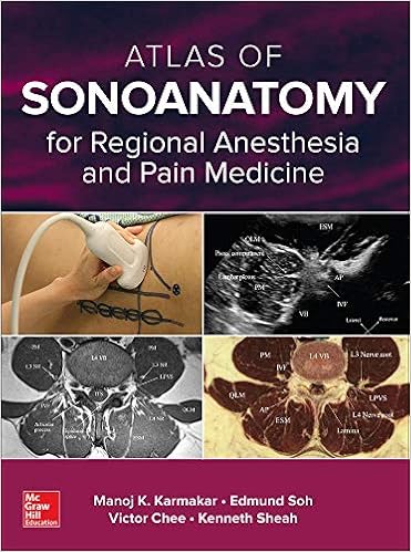
Atlas of Sonoanatomy for Regional Anesthesia and Pain Medicine
Hardcover – Illustrated, 2 December 2017
by Manoj Karmakar (Author)
A comprehensive full-color anatomical atlas designed specifically for the anesthesiologist and pain physician
A clear understanding of relevant anatomy is essential for physicians who wish to master ultrasound-guided nerve blocks. Although ultrasound images may help visualize the nerve to be blocked, the images still present a grainy and incomplete picture. Physicians looking to master US-guided nerve blocks are best served by becoming anatomical experts — and this innovative resource is an invaluable learning tool to doing just that.
In addition to 2D and 3D ultrasound images, Atlas of Sonoanatomy for Regional Anesthesia and Pain Medicine includes high-resolution CTs, MRIs, cadaver anatomy images, and anatomical illustrations to give physicians a comprehensive understanding of the anatomy of the neck, upper and lower extremity, trunk, thorax, thoracic spine, sacral spine, lumbar paravertebral region, and thoracic paravertebral region that are relevant to ultrasound-guided regional anesthesia.
Features
- Bulleted pearls impart how to obtain optimal ultrasound images at each site
- More than 600 full-color photographs and illustrations throughout
- Essential for anesthesiologists and pain physicians, and is also of value to musculoskeletal sonographers, radiologists, and emergency medicine physicians
- Enables you to learn and see, through full-color illustrations, the relevant anatomy for administering successful nerve blocks
- Includes CT and MRI correlations to ultrasound images, plus corresponding anatomical illustrations, cadaver anatomy, and photos of probe placement on the patient.
Although other texts may provide some of the imaging information contained in this unique atlas, this is the first resource to systematically and comprehensively gather all the imaging modalities for side-by-side comparison under one cover.
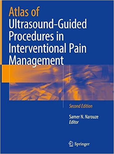
Atlas of Ultrasound-Guided Procedures in Interventional Pain Management
Hardcover – 30 May 2018
by Samer N. Narouze (Editor)
With a focus on anatomy and sonoantomy, this beautifully illustrated updated edition captures the latest advances in the rapidly growing field of ultrasound-guided pain medicine and MSK procedures. This atlas is divided into seven sections that provide an overview and focus on interventional approaches and advancements. Authored by international experts, each clinical chapter features a maximal number of instructive illustrations and sonograms and provides a description of sonoanatomy, instructions on performing the procedure and how to confirm appropriate needle placement. This book will help encourage and stimulate physicians to master approaches in interventional MSK and pain management.
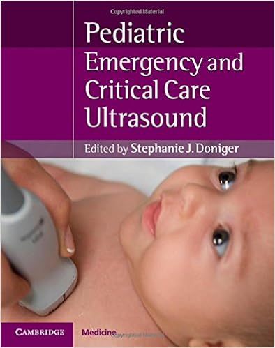
Pediatric Emergency Critical Care and Ultrasound
Hardcover – Illustrated, 24 April 2014
by Stephanie J. Doniger (Editor)
Emergency bedside ultrasound assessment is well established for adult patients, but has only recently been introduced into everyday clinical practice for the care of pediatric patients. Pediatric Emergency Critical Care and Ultrasound is a concise, practical text which explains the principles of ultrasound, its diagnostic application in all organ systems and its use as a procedural adjunct. Both well-established and innovative applications are described, assisting the practitioner in incorporating ultrasound into daily practice, facilitating patient care and decreasing radiation exposure. Case studies and abundant illustrations enable the reader to study the appropriate techniques in detail and learn from real examples from the pediatric emergency department and intensive care unit.
Pediatric Emergency Critical Care and Ultrasound is the first comprehensive bedside ultrasonography resource focusing on pediatric patients and is essential reading not only for pediatric emergency medicine subspecialists but for all emergency physicians, intensivists/critical care physicians and pediatricians.

Hardcover – 11 November 2021
by Harriet J. Paltiel (Editor), Edward Y. Lee (Editor)
This essential book is a unique, authoritative and clinically oriented text on pediatric ultrasound. It provides up-to-date information addressing all aspects of congenital and acquired disorders in children encountered in clinical practice. The easy-to-navigate text is divided into 20 chapters. Each chapter is organized to cover the latest ultrasound techniques, normal development and anatomy, anatomic variants, key clinical presentations, characteristic ultrasound imaging findings, differential diagnoses and relevant pitfalls. With more than 2400 images, examples of new technological developments such as contrast-enhanced ultrasound and elastography are included. Written by internationally known pediatric radiology experts and editorial team lead by acclaimed authors, Harriet J. Paltiel, MDCM and Edward Y. Lee, MD, MPH, this reference is a practical and ideal guide for radiologists, radiology trainees, ultrasound technologists as well as clinicians in other specialties with an interest in pediatric ultrasound.
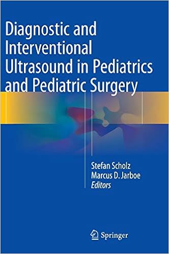
Diagnostic and Interventional Ultrasound in Pediatrics and Pediatric Surgery
Hardcover – 4 December 2015
by Stefan Scholz (Editor), Marcus D. Jarboe (Editor)
This book presents a comprehensive, state-of the-art guide and review of ultrasound applications for children and infants with surgical problems. It is meant as a single source to provide information about sonographic application, interpretation and technique for a diversity of pediatric surgical care providers, making it a useful tool for the ultrasound novice as well as the more advanced ultrasonographer. Sections address initial obstacles faced by a physician starting with ultrasound such as the scanning techniques, underlying anatomy and normal sonographic findings. The initial chapter provides an introduction and basic overview about ultrasound theory and techniques. Subsequent chapters focus on specific body parts and systems and their disease processes as it pertains to pediatric and neonatal patients. The text also includes a chapter on abdominal trauma and its evaluation with the FAST (focused abdominal sonography for trauma) exam.
Diagnostic and Interventional Ultrasound in Pediatric Surgery serves as a useful resource for a broad spectrum of pediatric care providers, including a growing number of ultrasound users, surgeons and pediatricians alike.
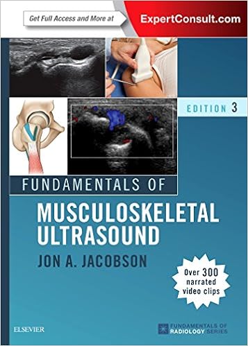
Fundamentals of Musculoskeletal Ultrasound
Paperback – Import, 11 September 2017
by Jon A. Jacobson MD (Author)
Effectively perform and interpret musculoskeletal ultrasound with this concise, highly illustrated resource by Jon A. Jacobson, MD. Fully revised, this bestselling title covers all the essential details of musculoskeletal ultrasound imaging, providing a solid understanding of the technique and how to make accurate diagnoses. It takes a concise, clear, and step-by-step approach to all of the most common musculoskeletal ultrasound applications, with specific details on anatomy, patient positioning, scanning techniques, normal and abnormal findings, tips, and pitfalls.
- A succinct, highly accessible writing style makes information easy to understand.
- Over 200 narrated video clips of real-time dynamic ultrasound imaging provide instruction in a succinct, didactic format, highlighting key findings.
- Common percutaneous ultrasound-guided musculoskeletal procedures are demonstrated, including transducer and needle positioning.
- Nearly 400 new ultrasound images show scanning technique, anatomy, and essential pathology.
- Over 200 narrated video clips of real-time dynamic ultrasound imaging provide instruction in a succinct, didactic format, highlighting key findings.
- Newly revised information throughout helps you grasp essential concepts in diagnostic musculoskeletal ultrasound, ultrasound-guided musculoskeletal procedures, and much more.
- Chapter 1, Introduction and Chapter 2, Basic Pathology Concepts now included in both print and electronic versions.
- Thoroughly revised text, references, and images keep you up to date.
- Expert Consult™ eBook version included with purchase. This enhanced eBook experience allows you to search all of the text, figures, Q&As, and references from the book on a variety of devices.
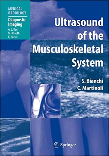
Ultrasound of the Musculoskeletal System
Hardcover – Illustrated, 5 March 2007
by Stefano Bianchi (Author), Carlo Martinoli (Author), A.L. Baert (Foreword), I.F. Abdelwahab (Introduction), L.E. Derchi (Contributor), G. Rizzatto (Contributor), M. Valle (Contributor), M.P. Zamorani (Contributor)
A comprehensive reference and practical guide on the technology and application of ultrasound to the musculoskeletal system. It is organized into two main sections. The first is devoted to general aspects, while the second provides a systematic overview of the applications of musculoskeletal ultrasound in different areas of the body. Ultrasound scans are correlated with drawings, photographs, images obtained using other modalities, and anatomic specimens. There is a generous complement of high-quality illustrations based on high-end equipment. This book will acquaint beginners with the basics of musculoskeletal ultrasound, while more advanced sonologists and sonographers will learn new skills, means of avoiding pitfalls, and ways of effectively relating the ultrasound study to the clinical background.
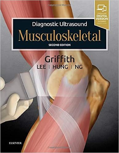
Diagnostic Ultrasound: Musculoskeletal
Hardcover – Illustrated, 19 January 2019
by James F. Griffith MD MRCP FRCR (Author)
Gain a solid understanding of musculoskeletal ultrasound anatomy, pathology, and technique with the second edition of this award-winning reference. Written by Dr. James F. Griffith and other leading experts in the field, Diagnostic Ultrasound: Musculoskeletal offers more than 100 detailed, clinically-oriented chapters of ultrasound anatomy, technique, diagnosis, differential diagnosis, reporting, and ultrasound-guided interventional procedures for the entire musculoskeletal system. This wealth of updated information helps you achieve an accurate musculoskeletal ultrasound diagnosis for every patient.
- Ensures that you stay on top of rapidly evolving musculoskeletal ultrasound practice and its expanding applications for everyday clinical use
- Contains new chapters on how to properly examine the joints of the upper and lower limbs with ultrasound and the best ultrasound technique for examining the groin, including groin herniae
Provides new information on ultrasound diagnostics and interventional techniques, keeping you up-to-date with improved accuracy of ultrasound diagnoses and clinical benefits of ultrasound-guided techniques, including joint injections for the upper and lower limbs - Uses a bulleted, templated format that helps you quickly find and understand complex information, as well as thousands of high-quality images and illustrations
- Describes how to write an efficient, useful, and factually correct ultrasound report
- Approaches musculoskeletal ultrasound from the viewpoints of a specific diagnosis (Dx section) as well as that of a specific ultrasound appearance (DDx section)
- Offers updates on fundamental ultrasound technique and ultrasound anatomy, ideal for those either new to musculoskeletal ultrasound or those with limited experience who wish to improve their skill
- An ideal reference for radiologists, sonographers, rheumatologists, orthopedic surgeons, sports physicians, and physiotherapists
- Expert Consult™ eBook version included with purchase, which allows you to search all of the text, figures, and references from the book on a variety of devices
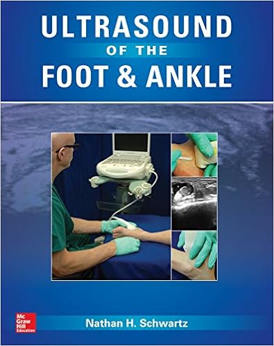
Ultrasound of the Foot and Ankle: Diagnostic and Interventional Applications
Hardcover – 2 December 2015
by Nathan Schwartz (Author)
Your definitive, step-by-step guide to ultrasound-guided exams and injections of the foot and ankle
Ultrasound of the Foot and Ankle is a multimedia, step-by-step introduction on how to use ultrasound successfully to diagnose and treat conditions of the foot and ankle. Authored by noted podiatric physician and educator, Nathan H. Schwartz, DPM, the book thoroughly covers both diagnostic and interventional methods.
Concise text and numerous illustrations provide you with easy to understand instruction on using ultrasound to accurately identify normal and abnormal anatomy of foot and ankle. In addition to an extensive section devoted to ultrasound-guided musculoskeletal injections, the book features chapters on ultrasound basics: image optimization, probe handling, and ergonomics. Numerous tips and tricks are provided to help you master exams and acquire new skills. It is the perfect introduction for practitioners who are eager to bring these procedures into their practices to enhance patient care.
- Guided learning that helps you put new procedures into practice
- Comprehensive coverage of how to effectively scan muscles, tendons, ligaments, joints, soft tissue, fat, bone, and nerves
- More than 60 videos illustrate key exams and injections
- Ideal for podiatrists, pain medicine specialists, sports medicine physicians, orthopedic surgeons, and physiatrists
Waldman’s Atlas of Diagnostic Ultrasound of Painful Foot and Ankle Conditions
Hardcover – 27 May 2016
by Waldman (Author)
With a focus on real-time clinical decision making, Dr. Waldman’s practical text helps you increase your diagnostic accuracy by performing dynamic scanning of the suspected pathology at the time the patient is being examined.
Derived from Waldman’s Comprehensive Atlas of Diagnostic Ultrasound of Painful Conditions, this unique resource helps guide your targeted point-of-care ultrasound examination of foot and ankle disease. Classic ultrasound images and anatomic drawings depict commonly encountered conditions, and Dr. Waldman’s expert advice guides you clearly toward the correct clinical diagnosis.
Key Features:
- An ideal reference for clinicians who would like to use, or are already using, ultrasonography to evaluate and treat foot and ankle disorders in their practice.
- Full-color line drawings, high-quality clinical photographs, and clearly labelled ultrasound images depict both common and uncommon pathologic conditions encountered in clinical practice.
- Chapters highlight anatomic considerations, physical diagnostic tests, clinical correlates, ultrasound techniques, clinical pearls, and more. \
- Written in the clear, concise, easy-to-read, “how to do it” style that is the hallmark of all of Dr. Waldman’s textbooks.
- A focus on the foot and ankle makes this a valuable resource podiatrists, orthopaedic surgeons specializing in foot and ankle, and other clinicians involved in the diagnosis and management of foot and ankle disorders.
Now with the print edition, enjoy the bundled interactive eBook edition, which can be downloaded to your tablet and smartphone or accessed online and includes features like:
- Complete content with enhanced navigation
- Powerful search tools and smart navigation cross-links that pull results from content in the book, your notes, and even the web
- Cross-linked pages, references, and more for easy navigation
- Highlighting tool for easier reference of key content throughout the text
- Ability to take and share notes with friends and colleagues
- Quick reference tabbing to save your favorite content for future use
Ultrasound Guided Musculoskeletal Procedures: A Practical Atlas
Paperback – 21 June 2021
by Sirisena, Dinesh (Khoo Teck Puat Hospital (KTPH) Sports Medicine Centre Singapore) (Author)
Ultrasound Guided Musculoskeletal Procedures in Sports Medicine: A Practical Atlas provides the support practitioners need based on practical, first-hand experience of a Sports and Exercise Medicine Physician who trained in musculoskeletal sonography. Over the years, and with much practice, the lessons learned and techniques developed are summarized with relevant pictures that guide those undertaking the procedure. As musculoskeletal ultrasound forms an important tool for physicians working in this field of medicine, this book helps physicians provide increasing expectation for patients who want a safe, guided procedure when clinically warranted. While an understanding of ultrasound imaging is essential prior to ultrasound guided procedures, there are few practical guides that provide practicing clinicians with a quick reference when faced with a procedure. This book fills that void.
Musculoskeletal Ultrasound Cross-Sectional Anatomy
Hardcover – Illustrated, 15 September 2016
by John C. Cianca (Author), Shounuck I. Patel (Author), Shonuck Cianca, John and Patel (Author)
This spectacular cross-sectional atlas provides a roadmap of normal sonographic anatomy of the musculoskeletal system with optimized images that emphasize spatial relationships and three-dimensional orientation. The book is designed to help novices acquire pattern recognition skills to resolve images into their anatomic components by pairing ultrasound scans with cross-sectional drawings. It will enhance familiarity with musculoskeletal anatomy as it appears on ultrasound imaging for practitioners at any level. Using a sectioned approach, the authors present a visual baseline for evaluating tendon, muscle, ligament, and nerve problems in the upper extremity, lower extremity, and cervical regions. Multiple high resolution views of each structure are accompanied by original illustrations that document the structures in the sonograph and serve as a reference to decipher the image and foster understanding of anatomic relationships and ultrasound appearances.
The atlas is an indispensable tool for clinicians learning diagnostic ultrasound, as they can use the anatomical images for comparisons with their own scans. For the seasoned practitioner, the organized format with high-resolution examples makes this an essential reference for confirming exam findings.
Key Features:
- Orients users to anatomical relationships best seen in cross section and necessary to effective utilization of diagnostic ultrasound
- Over 150 ultrasound images cover musculoskeletal anatomy from the shoulder to the foot
- Each ultrasound image has a correlative drawing in cross-sectional or regional format with the scanned area depicted within a highlighted frame to enhance understanding of the scanned anatomy
- Each image is accompanied by a body icon illustrating the level of the scan for each region
- Brief text points and legends emphasize key features and landmarks and offer technical tips for obtaining and interpreting scans.
Musculoskeletal Ultrasound in Rheumatology Review
Paperback – 5 October 2021
by Minna J. Kohler (Editor)
Point-of care ultrasound, or ultrasound performed and interpreted by the clinical in the clinic/office or at the bedside, has been rapidly expanding in use among multiple medical specialties. Musculoskeletal ultrasound has added value to clinical care to expedite diagnoses by visualizing mechanical versus inflammatory features and correlating these findings with patient’s symptoms.
The use of power Doppler to assess for subclinical inflammation or active hyperemia has been a distinguishing use of ultrasound for rheumatologists. Accurate needle guidance with ultrasound for joint and tendon procedures has allowed expansion of procedures in the clinic and has been associated with less pain and precise targeting of affected structures. Musculoskeletal ultrasound education is now offered in nearly all rheumatology fellowship training programs in the United States and is also included in residency training in other specialties (e.g. physical medicine and rehabilitation, orthopedics, podiatry, emergency medicine, and family medicine). Since the publication of the first edition of Musculoskeletal Ultrasound in Rheumatology Review, there has been tremendous growth of musculoskeletal ultrasound in the field of rheumatology as well as other musculoskeletal specialties.
The expanded second edition of this practical guide provides an updated clinical review of diagnostic and interventional applications of musculoskeletal ultrasound in rheumatology. New and revised chapters focus on the use of ultrasound in the diagnosis of specific rheumatic diseases such as osteoarthritis, scleroderma, psoriatic arthritis, gout, and lupus. Each chapter covers a standardized protocol of joint images with probe placement and includes numerous examples of common ultrasound pathologies. Study tools such as key-concept overviews, lists of important studies in the field, and extensive questions for self-assessment are included throughout.
Written by current experts and thought leaders in the rapidly advancing field of rheumatology ultrasound, the second edition of Musculoskeletal Ultrasound in Rheumatology Review is an essential reference for physicians and related professionals, residents, fellows, graduate students and nurses in rheumatology, imaging and radiology, sports medicine, internal medicine, and physiotherapy.
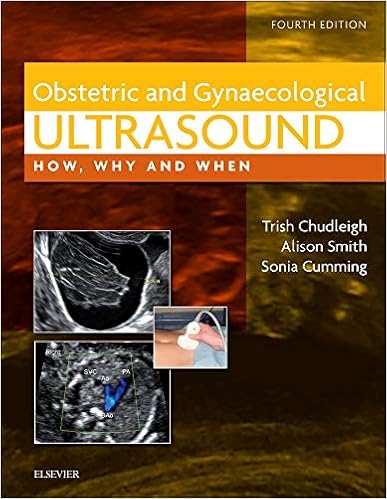
Obstetric & Gynaecological Ultrasound: How, Why and When
Hardcover – 20 September 2016
by Trish Chudleigh PhD DMU (Editor), Alison Smith MSc DMU (Editor), & 1 more
Obstetric and Gynaecological Ultrasound is a highly illustrated manual ideal for both the trainee and experienced sonographer that covers the full range of obstetric and gynaecological ultrasound examinations undertaken within a secondary referral setting. It combines the practicalities of how to perform these examinations with the information needed to interpret the findings and construct a clinically useful report.
The new edition of this well-established book expands its scope to provide an understanding of ultrasound imaging within the clinical management of the gynaecological patient. Elsewhere, its obstetric content has been fully updated with the latest technological, clinical and medico-legal information relevant to routine practice. Furthermore, its experienced sonographer authors continue to advise on the practical aspects of scanning as well as the role, value and limitations of ultrasound in the diagnosis of different diseases thus providing firm foundations in both obstetric and gynaecological imaging.
- Explains the principles of grey scale ultrasound, Doppler ultrasound and instrumentation
- Addresses problems from both practical and clinical viewpoints
- Provides comparative images showing results of good and bad scanning techniques
- Advises on how to communicate findings to a pregnant woman or gynaecological patient
- Discusses both the normal and abnormal ultrasound appearances for each of the relevant anatomical areas together
- Scope fully expanded to cover gynaecological ultrasound imaging including:
- the physiological changes taking place during the menstrual cycle
- the effects of exogenous hormones on the various ultrasound appearances during the menstrual cycle
- the ultrasound appearances of common abnormalities of the uterus, ovaries and adnexae
- the ultrasound assessment of an adnexal mass using a standardized approach
- New images to reflect the improvements in imaging technology
- New chapter on screening for Down’s syndrome and Edwards’ and Patau’s syndromes in accordance with current national screening recommendations
- New chapter on the medico-legal issues relevant to performing and reporting ultrasound examinations
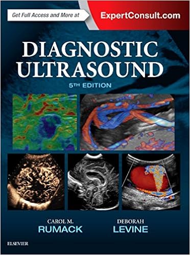
Diagnostic Ultrasound, 2-Volume Set
Hardcover – 10 October 2017
by Carol M. Rumack MD FACR (Author), Deborah Levine MD (Author)
Now fully updated with more than 2,000 new images, 200 new videos, and new content throughout, Diagnostic Ultrasound, 5th Edition, by Drs. Carol M. Rumack and Deborah Levine, remains the most comprehensive and authoritative ultrasound resource available. Spanning a wide range of medical specialties and practice settings, it provides complete, detailed information on the latest techniques for ultrasound imaging of the whole body; image-guided procedures; fetal, obstetric, and pediatric imaging; and much more. Up-to-date guidance from experts in the field keep you abreast of expanding applications of this versatile imaging modality and help you understand the “how” and “why” of ultrasound use and interpretation.
- Covers all aspects of diagnostic ultrasound with sections for Physics; Abdominal, Pelvic, Small Parts, Vascular, Obstetric, and Pediatric Sonography.
- Uses a straightforward writing style and extensive image panels with correlative findings.
- Features 5,000 images – more than 2,000 brand-new – including new 2D and 3D imaging as well as the use of contrast agents and elastography.
- Includes a new virtual chapter on artifacts with individually labelled images from throughout the book, displaying artifacts with descriptive legends by category and how they can be used in diagnosis or corrected for better quality imaging.
- Features more images and new uses for contrast agents in the liver, breast, and in pediatric applications. Includes current information on imaging more diagnostic dilemmas, such as Zika virus in the fetus and newborn.
- Includes 400 video clips showing real-time scanning of anatomy and pathology.
- Expert Consult™ eBook version included with purchase. This enhanced eBook experience allows you to search all of the text, figures, Q&As, and references from the book on a variety of devices.
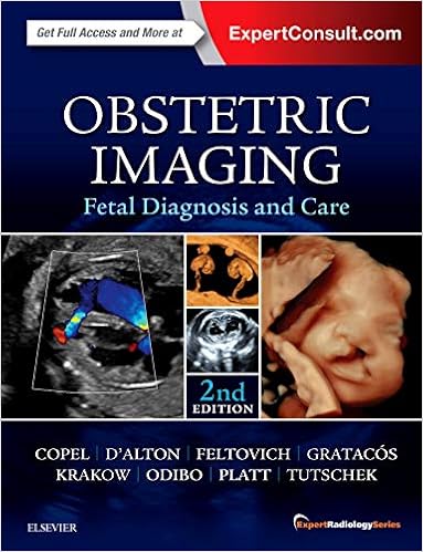
Obstetric Imaging: Fetal Diagnosis and Care: Expert Radiology Series
Hardcover – Illustrated, 15 September 2017
by Joshua Copel MD (Author), Mary E. D’Alton MD (Editor), Helen Feltovich (Editor), & 4 more
Richly illustrated and comprehensive in scope, Obstetric Imaging, 2nd Edition, provides up-to-date, authoritative guidelines for more than 200 obstetric conditions and procedures, keeping you at the forefront of this fast-changing field. This highly regarded reference covers the extensive and ongoing advances in maternal and fetal imaging in a concise, newly streamlined format for quicker access to common and uncommon findings. Detailed, expert guidance, accompanied by superb, high-quality images, helps you make the most of new technologies and advances in obstetric imaging.
- Features more than 1,350 high-quality images, including 400 in color, and over 85 videos online.
- Helps you select the best imaging approaches and effectively interpret your findings with a highly templated, bulleted, at-a-glance organization.
- Reflects all the latest developments in the field, including genetics, open fetal surgery, fetal echocardiography, Zika virus, and 3D imaging, so you can provide the safest and most responsive care to both mother and fetus.
- Includes new chapters on Limbs and Bones Overview; Open Fetal Surgery; Biophysical Profile; Ultrasound Physics; Elastography; Doppler; MRI; Echogenic Bowel; Pregnancy of Unknown Location (PUL), Failed Pregnancy and Ectopic Pregnancy, Cesarean Scar Pregnancy; Cytomegalovirus (CMG), Rubella, Toxoplasmosis, Herpes, Varicella; and Congenital Syphilis; plus a new chapter on Zika Virus written by imaging experts from the “hot zone.”
- Keeps you up to date with the latest developments in multimodality imaging and optimizing diagnostic accuracy from ultrasound, 3D ultrasound, Doppler, MRI, elastography, image-guided interventions, and much more.
- Expert Consult™ eBook version included with purchase. This enhanced eBook experience allows you to search all of the text, figures, Q&As, and references from the book on a variety of devices.
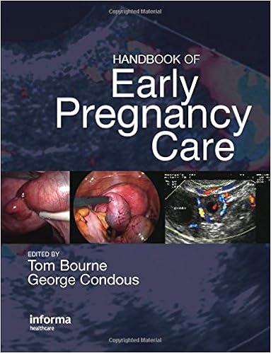
Handbook of Early Pregnancy Care
Hardcover – Illustrated, 17 October 2006
by Thomas H. Bourne (Editor), George Condous (Editor)
Problems in early pregnancy are among the most common conditions in gynecology and – with management becoming less invasive and depending more on accurate ultrasound and a good understanding of serum biochemistry – many countries now recommend that all maternity units have an early pregnancy section dedicated to managing these conditions.
In addition to this, the subject is also an essential part of the training syllabus in the speciality of maternal-fetal medicine. Handbook of Early Pregnancy Care is a practical guide for the management of at-risk early pregnancy.
The chapters are concise, well-illustrated and contain decision trees for the management of each condition. Focusing on practical management, not just research, this helpful text will be beneficial in the clinic, as contributors are encouraged to tell the readers what to do in critical situations. Covering key points and containing illustrative case studies, this handbook is relevant to trainees in maternal-fetal medicine and obstetrics, as well as nurses and sonographers who see many at-risk patients in the early stages of pregnancy.
Ultrasound in Subfertility: Routine Applications and Diagnostic Challenges
Hardcover – 1 August 2018
by K. (Author), M.D. (Author), Ph.D. Jayaprakasan (Author), Sonal (Author), M.D. Panchal (Author)
The second edition of this comprehensive reference provides practitioners with the latest advances in the use of ultrasound for diagnosis and management of subfertility.
Divided into thirteen chapters, the book begins with an introduction to the principles and applications of ultrasound, and examination techniques and in pelvic assessment.
The next sections cover the use of ultrasound for the diagnosis of different gynaecological conditions that may affect fertility, followed by ultrasound-guided procedures in assisted reproduction and potential complications.
The text concludes with chapters on ultrasound in male infertility, and three-dimensional ultrasound in subfertility.
Edited by recognised experts in the field, the text is further enhanced by more than 400 ultrasound images and is accompanied by an interactive DVD ROM providing video clips for each clinical scenario described in the book.
Key points
- Second edition presenting latest advances in use of ultrasound in subfertility
- Includes DVD ROM of video clips demonstrating clinical scenarios
- Internationally recognised editor team
- Previous edition (9789351520108) published in 2014
Ultrasound For Advanced Practitioners In Pregnancy And Women’s Health
Paperback – Illustrated, 11 February 2019
by Cydney Afriat Menihan (Author)
Each new print copy includes Navigate 2 Preferred Access that unlocks a comprehensive and interactive eBook and Navigate 2 TestPrep.
Ultrasound for Advanced Practitioners in Pregnancy and Women’s Health is an indispensable resource for midwives, OB/GYN nurse practitioners, OB/GYN physician assistants, and students. A unique text, it addresses specific clinical application of ultrasound technology in all aspects of women’s health care. Developed by an advanced practitioner for advanced practitioners, this resource includes the basics of ultrasound technology, step-by-step illustrations, and review questions.
Ultrasound for Advanced Practitioners in Pregnancy and Women’s Health is also an ideal reference to prepare for the newly developed Midwife Sonography Certificate administered by the American Registry for Diagnostic Medical Sonography (ARDMS).
How to Perform Ultrasonography in Endometriosis
Hardcover – 25 October 2018
by Stefano Guerriero (Editor), George Condous (Editor), Juan Luis Alcázar (Editor)
This structured dynamic book outlines, step by step, an evidence-based systematic approach to the sonographic evaluation of the pelvis in women with suspected endometriosis. This “how to” guide is intended for those with basic ultrasonography skills who want to further develop their capabilities in performing the relevant sonographic techniques to identify endometriosis. Detailed schematics, and corresponding high-resolution ultrasound images and intuitive videos support readers in expanding their technical skills and bridging the gaps in their knowledge of endometriosis ultrasound. The International Deep Endometriosis Analysis (IDEA) group consensus statement was the culmination of the work of 29 authors from 5 continents. With the publication of How to Perform Ultrasonography in Endometriosis the authors intend to provide the basis for quality improvement and benchmarking of ultrasound in the world of endometriosis.
This book not only offers sonologists, radiologists and sonographers valuable insights into the field of endometriosis ultrasound, but also enables them to develop their practical skills in assessing women with chronic pelvic pain.
Guidelines for Nurse Practitioners in Ambulatory Obstetric Settings 2ed
Paperback – 15 May 2017
by Nancy J. Cibulka (Author), Mary Lee Barron (Author), Mary Cibulka, Nancy and Barron (Author)
The only comprehensive source of current, evidence-based guidelines for nurse practitioners, nurse-midwives, clinical nurse specialists, and other health professionals who provide prenatal and postpartum care in outpatient settings, this clinical reference covers the latest health care guidelines in an easy-to-read, outline format. With three new chapters, Obesity in Pregnancy, Dermatological Concerns, and Preventing Zika in Pregnancy, the second edition has also been updated to include new guidelines from professional organizations such as the Association of Women’s Health, Obstetric and Neonatal Nurses; American Congress of Obstetricians and Gynecologists; American Academy of Pediatricians; and the U.S. Preventive Services Task Force.
Delivered in a convenient, spiral-bound format for health professionals in a busy clinical practice setting, this guide addresses clinical topics and practice standards regarding preconception, prenatal, and postpartum nursing care. Topics are broken down into definition, etiology, history, physical exam, lab exam, differential diagnosis, treatment, complications, consultation/referral, and follow-up. Sections provide in-depth detail on genetic counseling, health promotion and assessment, lactation issues, and medications during pregnancy. Tables and diagrams further illustrate and reinforce the content, and numerous web addresses are provided.
New to the Second Edition:
- Three new chapters, Obesity in Pregnancy, Dermatological Concerns, and Preventing Zika in Pregnancy
- New and updated guidelines from the Association of Women’s Health, Obstetric and Neonatal Nurses; American Congress of Obstetricians and Gynecologists; American Academy of Pediatricians; and the U.S. Preventive Services Task Force
- New clinical updates on genetic screening and testing, nausea and vomiting, use of low-dose aspirin to prevent pregnancy complications, HIV prevention using PrEP, breastfeeding, avoidance of nonmedically indicated early-term delivery, and use of marijuana and heroin
- New nomenclature for preeclampsia
- Updated information on healthy preconception care
Key Features:
- Comprises the only comprehensive source of current guidelines for nurse practitioner care of obstetric patients in ambulatory settings
- Presents complete guidelines in consistent, easy-to-access outline format
- Written by board-certified nurse practitioners with more than 50 years of combined women’s health and obstetric/perinatal practice
- Offers guidelines on identifying complications and when to refer for specialist care
Paperback – 13 February 2018
by Vivek S. Tayal (Editor), Michael Blaivas (Editor), Troy R Foster (Editor)
This book addresses the wide range of issues that face the program leader – from how to choose a site and how to negotiate for equipment, to how to determine staffing requirements and how to anticipate and defuse possible turf issues with other programs and services in the hospital or healthcare facility.
The early chapters of this book focus on the leadership of your program whether in your department or institution. The second section centers on education at all levels recognizing that smaller machines have made ultrasound available for medical students to advanced practitioners. The third section provides detailed logistics on equipment, maintenance, and safety. The fourth section focuses on a quality improvement program and includes a chapter on the workflow process. For those with limited budgets we also offer a section on practical operating and educational solutions. The fifth section offers insight into hospital level credentialing, quality assurance, national politics, and recent issues with accreditation. This is followed by reimbursement and coding. The last section covers topics in specialized communities.
Chapters focus on ultrasound in global health, emergency medical services, pediatrics, critical care, community and office based practices. Multiple US working documents including checklists, graphs, spreadsheets, tables, and policy appendices are included.