Welcome to the POCUS Educator / SEED resource page
If you’re a qualified sonographer looking to break from the radiology world, want to utilise the skills you’ve spent so long developing, but are not quite sure of the options available to you then look no further.
Congratulations – you’ve taken the first steps toward uncovering the complexity and wonder associated with transitioning to the world of ultrasound education at the point-of-care.
We’ve developed this resource as a one-stop shop to give you some insight into what it takes to become an educator in the POCUS (point-of-care ultrasound) world.
While it’s been created with a distinctly Australasian flavour, we’ve taken some of what we’ve learned from our decades of teaching POCUS users around the world and collated information that is relevant to anyone looking to transition from a clinical ultrasound role to a role where the scope of practice extends far beyond imaging.
Be sure – we make no bones about the fact that transitioning to a POCUS educator role isn’t for everyone. If you can scan quickly and efficiently then you possess about 60% of the skills you need; the role requires investment in the development of a range of new skills and abilities that will challenge at a personal and professional level (see Suean’s video).
However the explosion of the use of ultrasound at the point of care means that becoming a POCUS educator should now be legitimately regarded as a new specialist career pathway, a rewarding choice that can take you places. Heck – it’s what we’ve been doing for the past two decades.
We intend this resource to remains a living page, and anticipate that it change as the world develops.
We’d love for you to contribute; if you have a tip, a trick or a resource you think others would benefit from let us know.
And join our POCUS educators/SEED Support Network Facebook group – we’re starting this up as a place we can share ideas in a safe environment.
Get on board.
Suean, Mike and the Zedu Crew
Join our POCUS educators/SEED Support Network Facebook group

What does it take to become a
POCUS Educator/SEED
In this edition of the monthly Coaching Corner, Suean and friends discuss what it means and what it takes to be a SEED – Sonographer Educator in the Emergency Department.
While the SEED is a uniquely Australian construct focused on the emergency medicine context, the information is relevant to anyone wanting to make the transition to being a POCUS educator.
In this video you’ll learn what a SEED is, what the role involves and the type of ultrasound examinations you’ll need to master in order to teach others. Hear what the differences are between ‘traditional’ radiology ultrasound and Emergency Department ultrasound. Understand the steps you will need to take to make the transition from Sonographer to Sonographer Educator in the ED.
Want to join us on the Coaching Corner free each month? Register here…
Join us at TRain the UltraSound Trainer (TRUST™) – THE ultimate ultrasound instructor’s course
Don’t have enough time to develop your student’s skills, let alone your own teaching skills? Do you want to be a better teacher but don’t know where to start? Are you frustrated your learning intentions are not translating to the learning outcomes you need fast enough?
Effective ultrasound teaching requires more than knowing how to scan. Zedu’s TRain the UltraSound Trainer (TRUST™) course is designed to transition you from competent scanner to confident educator.
The TRUST™ course is THE ultimate ultrasound instructor’s course that will give you the shortcuts to upskill your learners quickly.
This program is designed to give you the skills and knowledge to help you TEACH better and accelerate your student’s progress.
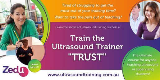
Learn how to structure your ultrasound teaching to make learning easy for your learner, and life simpler for you.
Internationally recognised, this course is highly recommended for anyone who is passionate about becoming the best ultrasound instructor they can be.
But don’t just take our word for it…check out what others have to say.
While point-of-care ultrasound (POCUS) is within the scope of practice for all medical professionals, it has (rightly or wrongly) become synonymous with emergency medicine. This is primarily due to the fact that emergency physicians based in the USA have for the past 20 plus years been at the forefront of exploring the utility of the technology across the full spectrum of cases they are presented.
Yet while the ED fraternity has been most vociferous in their advocacy of ultrasound (take their dominance on the topic in the social media space), other specialties should not be forgotten.
Bedside ultrasound has been a significant part of anaesthesia, O&G, surgery, intensive/critical care and general medicine for decades, with other physician specialists recognising the benefits ultrasound can bring for rapid diagnosis and treatment. In outback Australia POCUS has proven its utility for our rural generalist friends. POCUS in pre-hospital medicine in Australia is gaining traction. Nurses and midwives are increasingly expected to upskill to provide the best in patient care, and allied health professionals such as podiatrists and physiotherapists are seeing the immediate benefits imaging an injury can deliver for rehabilitation.
So – while the content of this page also is emergency-centric in nature – we musn’t forget that POCUS educators have a role to play in all areas of medicine. Which one best suits you?
|
eFAST
Synonymous with point of care ultrasound, the eFAST scan forms the mainstay of the emergency medicine ultrasound armoury. Originally confined to a 3 view FAST (Focused Abdominal Sonogram for Trauma) exam to rule out abdominal bleeding due to trauma (right upper quadrant, left upper quadrant, pelvis), the approach has extended over time to include imaging of the heart and lungs to become the eFAST (Extended Focused Assessment with Sonography in Trauma).
The eFAST Exam is designed to provide answers to 4 binary questions in haemodynamically unstable patients: is there a pneumothorax? Is there fluid in the pericardium? Is there free fluid in the abdomen? Is there free fluid in the thorax?
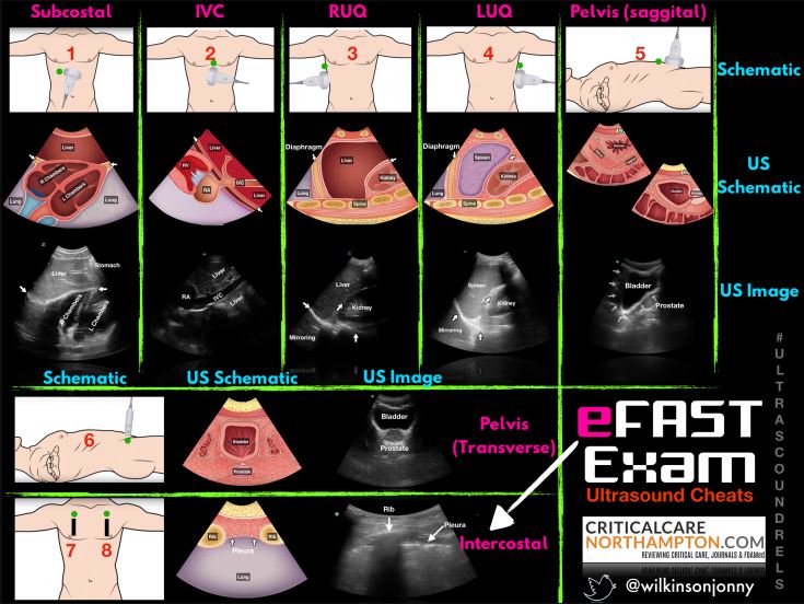
Image: courtesy Jonny Wilkinson
The rapid implementation of the eFAST protocol requires good hand eye co-ordination, image optimisation skills (both window acquisition and system optimisation) and diagnostic pattern recognition. It also requires a discipline to maintain a binary ‘Yes’/ ‘No’ approach to answering the clinical questions, and the ability to call it quits when it’s clear good imaging (diagnostically acceptable) is not possible.
If you’re a POCUS educator in the ED, you’ll need to become intimately familiar with the eFAST scan and extend your skills to include cardiac and lung imaging. Even if you’re not in the ED or an emergent setting, the imaging protocols of eFAST are applicable in other clinical settings. Fundamental to your role as a POCUS educator, you’ll need to ensure that your learners develop good probe handling skills, pattern recognition and stay focused. From your point of view, you’ll also have to make the transition to a point of care mindest, as the approach to imaging differs significantly from that of a traditional radiological setting. Our analogy is you are moving way from being a photojournalist with the time and space to compose your images, and towards being a tourist taking the snapshots you’ll need to prove you’ve been and seen.
If you get this you’re on your way.
For guidance on eFAST check out any of the iBook or other resources we have listed below.
Cardiac
Whether performed as part of the eFAST protocol due to trauma or due to other indications (hypotension, suspected pulmonary embolism, acute heart failure, syncope, chest pain or dyspnea, cardiac arrest, or assessment of volume responsiveness), a focused cardiac ultrasound can provide significant – potentially life saving – information.
As a POCUS educator you don’t have to become a fully blown echocardiographer to get utility out of heart imaging. More often than not there isn’t the luxury of time to take precise cardiac measurements. But what you will need is the ability to get a good B-mode image, develop and teach good pattern recognition skills using a 5E style approach – including eyeballing poor cardiac function – and identify ways to teach image acquisition in suboptimal conditions.
|
You’ll need to get familiar with the cardinal imaging series – parasternal (short-axis |PSAX, long-axis | PLAX), apical (4 chamber | AP4, 5 chamber | AP5, coronary sinus), subcostal, and inferior vena cava (IVC).
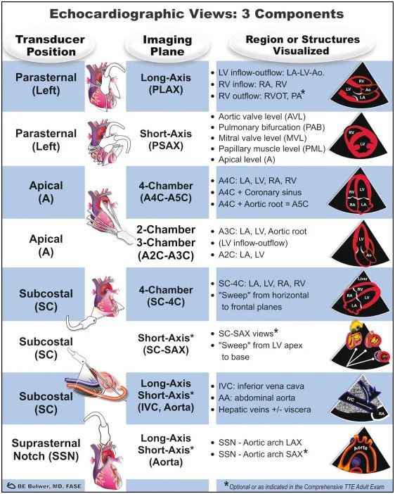
Image: courtesy Bernard E. Bulwer, MD, FASE.
In Australia you’ll also have to deal with the issues associated with probe marker orientation – accommodating the cardiology convention that it appears on the right hand side of the image.
|
For guidance on cardiac ultrasound at the point of care, check out any of the iBook or other resources we have listed below.
Lung
Once upon a time – not all that long ago – lung ultrasound was considered impossible, air being the enemy of soundwaves. But as with most things, someone saw a pattern in what wasn’t visible and determined that lung ultrasound was not only possible, but clinically valuable.
|
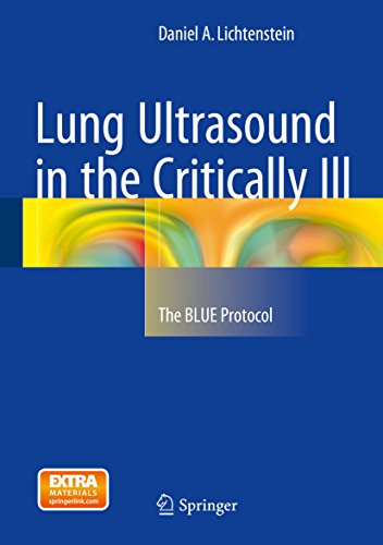
While there’s debate around who was first to ‘discover’ the utility of lung ultrasound, the individual credited with protocolising its use is Daniel A. Lichtenstein – a Paris based intensivist. He has been working in the field since the early 90’s, first pioneering work on lung sliding for pneumothorax, and is famous for continuing to use a dinosaur of an ultrasound system! His most notable work is the creation of two protocols adapted from lung ultrasound: the bedside lung ultrasound in emergency (BLUE)-protocol for the immediate diagnosis of acute respiratory failure and the fluid administration limited by lung sonography (FALLS)-protocol for the management of acute circulatory failure.
Dr Lichtenstein has created a unique nomenclature (read non traditional that will confuse sonographers) that indicates he was not traditionally schooled in ultrasound physics, but his pattern recognition means we can now use images generated by the interation of air and fluid to diagnose illness. His contribution is priceless, and his work has informed a legion of physicians globally that has paid dividends during covid.
And if you’d like an insight into the man who is Daniel Lichtenstein check out the presentation he gave at the SMACC conference in 2017 – Lung Ultrasound in Critical Care and Resuscitation.
Want more?
You can purchase Daniel Lichtenstein’s book on Amazon
Abdominal Aortic Aneurysm (AAA) & Aortic Dissection
Abdominal Aortic Aneurysm (AAA) & aortic dissection are vascular emergencies that POCUS was made for; with >90% sensitivity for detecting AAAs it is the investigation of choice in the unstable patient and may improve time to diagnosis.
As a POCUS educator or SEED this is the examination that will be most familiar. However – unlike the radiology setting where you are following a screening protocol – in the emergent or point of care context the patient will be presenting in acute pain and will need urgent treatment.
Your job will be to make sure that those under your tutelage are measuring the right structure. The errors that are made are different to those in the radiology context. Where you may be concerned at the accuracy of your measurement, physicians make different errors; the errors in their world are at the broader level, and it is not uncommon for them to measure the SMA or even the vertebral body. On this basis your job is to make sure the technique is correct – that the depth setting is right, ensuring they’re imaging perpendicular and scanning proximally, and can work around bowel gas.
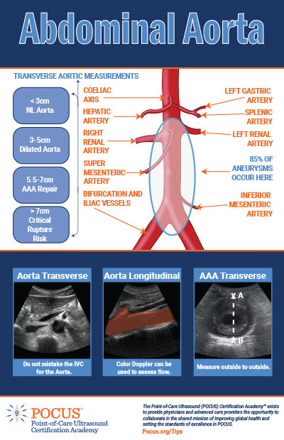
Image: courtesy The Pocus Certification Academy
Again, you’ll need to resist the sonographer’s temptation to get the perfect image, and instead focus on ensuring that your learners get a clinically acceptable image that can answer the question ‘Is there a AAA/dissection’ with a binary ‘Yes’/’No’ answer.
Check out the pitfalls of scanning the aorta from Suean’s very first Coaching Corner at the start of covid lockdowns – where we were all learning how to use the technology!
Procedural guidance
Ultrasound assisted procedural guidance improves the success rate of procedures, while decreasing complications. Whether it’s a relatively simple peripheral IV insertion, central line placement, a foreign body location, or at the more extreme end a pericardiocentesis.
|
However needle guidance with ultrasound is a core skill whose difficulty is often underrated, and as a POCUS educator/SEED your focus will be on ensuring your learner has the skill and ability to guide a needle in- and out-of-plane – regardless the application.
Get your preparation, set up and alignment right and you’ll hit your target every time. If you’re looking for guidance for your learners check out this edition of Suean’s Coaching Corner which focuses on what are some of the problems you may encounter with needle guidance and why.
|
Who doesn’t love something for free – right? And when that something is free, online, high quality and all about ultrasound then count us in!
We’ve listed a small number of ‘freebies’ that – if you want to become a POCUS educator or SEED – you should not go without. Click on the links and you’ll download immediately. The ones listed here are all built for planet iOS (Apple) – which is the majority – but there are a few pdf resources available too. Click here for our full list of downloadble free books – from anaesthesia to O&G here’s something for every POCUS aficionado.
Introduction to Bedside Ultrasound – part 1 [iBook]
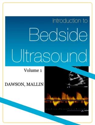
Introduction to Bedside Ultrasound seeks to teach bedside ultrasound to students in a more efficient, directed format than previous texts. It accomplishes this with a plethora of multimedia adjuncts. Instead of simply reading about ultrasound pathology and protocols, the reader can watch actual videos and demonstrations of the pathology from directly within the book while reading. This experience is closer to bedside learning with a true expert than reading a traditional textbook.
Rapid Ultrasound: Basics & RUSH exam [iBook]
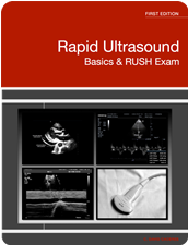
This book introduces the clinician in training to the ultrasound machine and its uses for diagnostic purposes in the critical care setting.
In addition, it helps students familiarize with the ultrasound machine knobs, probes and the RUSH protocol.
Introduction to Bedside Ultrasound – part 2 [iBook]
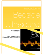
Introduction to Bedside Ultrasound Volume 2 like the 5 star rated volume 1 teaches bedside ultrasound in a more efficient, directed format than previous texts. It accomplishes this with a plethora of multimedia adjuncts. Instead of simply reading about ultrasound pathology and protocols, the reader watches actual videos and demonstrations of the pathology from directly within the book while reading. Volume 1 has received incredibly rave reviews, but Volume 2 is as good or better with more content, advanced techniques, more video, and more demonstrations. The experience is closer to bedside learning with a true expert than reading a traditional textbook.
Trauma Ultrasound eBook [iBook]

The use of ultrasonography revolutionized trauma care, and now this one-of-a-kind interactive eBook is going to revolutionize trauma ultrasound education. Written and produced by nationally known emergency medicine educators and sponsored by the American College of Emergency Physicians. Seven chapters, more than 40 full-motion videos, and more than 50 diagnostic images, photographs, and illustrations, many of which are interactive to enhance the learning experience.
We’re regularly contacted for our recommendations on books and other resources that we think are valuable to further ultrasound knowledge.
Whatever the topic, we’ve got our favourites – you can’t beat a good old fashioned book. Click here for a full list of our top picks – from musculoskeltal to obstetrics we’ve got it covered. And a big thanks to you as each purchase through this webpage helps support us through affiliate partnerships.
Emergency Ultrasound Made Easy
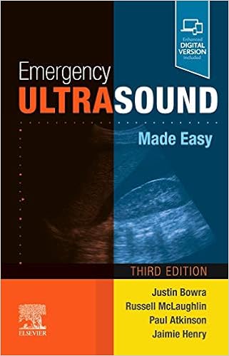
by Justin Bowra MBBS FACEM CCPU (Editor), Russell McLaughlin MB BCH BAO FRCSI MMedSci FCEM CFEU (Editor), Paul Atkinson MB BCh BAO (Hons) MA FRCEM FRCPC CFEU (Editor), Jaimie L Henry MBBS BSc (Hons) MRCP (Editor)
This simple, jargon-free text fits in your pocket, providing an ‘on-the-spot’ guide to clinician-performed ultrasound in the emergency department, intensive care unit or in the field.
Written by an international team of experts and comprehensively updated in its third edition, Emergency Ultrasound Made Easy brings together in one volume the latest indications for focused ultrasound, including those related to the COVID-19 pandemic.
The text is highly accessible and easy to use in an emergency. It is aimed at the rapidly expanding cohort of non-radiologist clinical sonographers who use focused ultrasound. However, its broad scope (for example using ultrasound in the rapid diagnosis of DVT) makes it an invaluable addition to the library of any doctor with an interest in the technique, whether in primary care or the hospital setting.
The Ultimate Guide to Point-of-Care Ultrasound-Guided Procedures
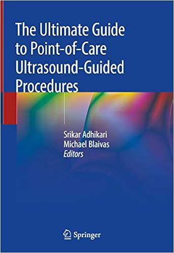
by Srikar Adhikari (Editor), Michael Blaivas (Editor)
This comprehensive book provides an in-depth examination of a broad range of procedures that benefit from ultrasound guidance in the point-of-care setting. It covers common procedures such as ultrasound-guided central and peripheral venous access to regional nerve blocks, temporary pacemaker placement, joint aspirations, percutaneous drainage, a variety of injections and airway management.
Chapters examine a variety of topics critical to successful ultrasound procedures, including relevant sonoantomy, necessary equipment, proper preparation, potential complications, existing evidence and how to integrate these procedures into clinical practice. For each procedure, the book includes step-by-step instructions and discusses the advantages of ultrasound guidance over traditional techniques.
Providing rich procedural detail to help in clinical decision making, The Ultimate Guide to Point-of-Care Ultrasound-Guided Procedures is an indispensable, go-to reference for all health care providers who work in a variety of clinical settings including primary care, emergency department, urgent care, intensive care units, pediatrics, pre-hospital settings and those who practice in the growing number of new ultrasound programs in these specialties.
If you’re going to be a POCUS educator or SEED, then it is inevitable that you will have to design and deliver presentations. And we all know what makes a bad presentation – we’ve all suffered through them. But what does it take to make a great presentation, and what tools are available?
We have our own approach to presentations here at Zedu, developed over the years. We base our presentations on imagery and story telling in a short format – employing visual cues to relate information. But what works for us may not work for you. What you need to do is find a voice and style that you’re comfortable with. To get good you must practice, and get to know your material intimately. There’s no better way to learn than to teach. And don’t forget to become intimate with your technology – nothing derails a presentation quicker than an IT meltdown.
Check out the following websites for ways you can improve your presentations.

p cubed presentations – the brainchild of Ross Fisher – is a concept about presentations and how to improve them. The aim is to share ideas about presentations, their construction, design and delivery such that the message is not lost, in time, like tears in rain. Ross’ mission is to help presenters realise their goal of effective presentations, stimulate debate and value effective communication.
A downloadable tool designed by Ben Smith to batch anonymize ultrasound clips: ClipDeidentifier. Ideal for inserting clips into your presentations without the information that could identify the patient.

Nancy Duarte is author of six best-selling books and CEO of Duarte, Inc. Duarte is a communication expert who incorporates story patterns into business communications.
Check out her ‘Resource Hub’, which provides a numer of resources you can use to brush up on your communication skills – long reads, quick tips, or tools.
In the POCUS space, a qualified physician is considered appropriately skilled and able to interpret – if not use – ultrasound; it is deemed to be within scope of practice.
However, while unfettered utilisation of ultrasound may be theoretically feasible, many workplaces determine the standards that must be attained by an individual to gain usage privileges in a clinical context. This is most advanced in the hospital context, where it is not unusual for local health authorities to defer to the relevant ultrasound credentialing standards established by the college under which a physician falls. Bodies external to employers and Colleges do offer forms of credential that recognise POCUS skills which can be of benefit.
As a POCUS educator/SEED it’s important that you become familiar with the colleges that create the rules of ultrasound use, as they ultimately determine what your learners (and you) can and can’t do. The reality is that guidelines, statements and protocols create the ‘rules of engagement’ that inevtably have unavoidable implications. You must also be aware of the credentialing options available, and the associated processes/costs involved, so that you can guide and assist your learners in the best way possible.
Local Health Authorities/Districts
Local Health Authorities/Districts determine the ultrasound credentialing standards of users under their employ. This by default typically references the standards established by the Colleges under which their medical professionals fall. You can find credentialing standards for ultrasound articulated by the following colleges, supported variously by subcommittees, special interest groups and affiliated organisations/societies:
- The Australasian College for Emergency Medicine (ACEM)
- Internal committee – ACEM Emergency Department Ultrasound Committee (EDUC)
- Affiliated external interest group – Emergency Medicine Ultrasound Group (EMUGs)
- The Australian and New Zealand College of Anaesthetists (ANZCA)
- Internal committee – Anaesthesia Continuing Education (ACE) Committee is a tripartite body governed by ANZCA, Australian Society of Anaesthetists (ASA) and New Zealand Society of Anaesthetists (NZSA).
- Affiliated external society – Australian Society of Anaesthetists (ASA)
- The Australian College of Rural and Remote Medicine (ACRRM)
- The College of Intensive Care Medicine Australia and New Zealand (CICM)
- Internal SIG – Critical Care Ultrasonography Special Interest Group (CICM)
- The Royal Australasian College of Physicians (RACP)
- There are 51 Specialty Societies affiliated with the RACP, many with their own ultrasound frameworks.
- The Royal Australasian College of Surgeons (RACS)
- The Royal Australian and New Zealand College of Obstetricians and Gynaecologists (RANZCOG)
- Internal committee – Obstetrical & Gynaecological Ultrasound (COGU) Subspecialty Committee
- The Royal Australian College of General Practitioners (RACGP)

The Point-of-Care Ultrasound (POCUS) Certification Academy offers globally recognized certifications for healthcare providers to independently validate their POCUS knowledge, application, and proficiency. Check out their POCUS certification options which meet and exceed Australian benchmarks.
Australasian Society for Ultrasound in Medicine (ASUM)
ASUM (not to be confused with ACEM) offers a variety of ultrasound credential pathways for a range for specialist clinicans and allied health professionals.
Physicians can seek to qualify for any one of 26 Certificates in Clinician Performed Ultrasound (CCPU) of the more advanced Diploma in Diagnostic Ultrasound (DDU). Similarly, allied health professionals can seek to qualify for any one of 11 Certificates in Allied Health Performed Ultrasound (CAHPU). Check out their website for more information.
So you’ve made the decision to pursue a career as a POCUS educator/SEED. Now what?
If you’ve secured a stable position as a hospital employee congratulations – you’ve hit the jackpot. However, not all hospitals or medical facilities have the resources to maintain a fully funded POCUS educator/SEED position, so the majority of you will likely find yourself operating as an independent agent. This will essentially mean you’ll be starting your own business.
Before you take the leap, please take the time to set yourself up in a manner that best meets your requirements. While we’re not a financial adviser, it’s vitally important that you have the structures and processes in place to ensure a) you get paid and b) meet your obligations. Don’t underestimate how much time you’ll need to dedicate to administration – even if it is a business of one.
Starting your own business
![]()
If you’re starting out as a POCUS educator, you may wish to start a business.
While we’re not business advisers, there are tax and super responsibilities you need to be aware of, including:
- the tax implications of your business structure
- whether you’re entitled to an ABN
- registering your business
- records you need to keep
- deductions you can claim.
Read all about what you need to know and do when starting your own business on the Australian Taxation Office website.
Administration tools
While you can keep your own spreadsheets to track your finances, to keep the tax man happy we recommend that you use a reputable financial management/accounting software provider.
The major providers in the Australian market are:
Each offer slightly different packages and pricing models, so it’s worth doing your homework to see what will work for you. Also, consult your accountant – working with a system they are familiar with makes life a lot easier.
Last, but not least, is the icing on the cake – social media and useful websites.
On the social media front, we recommend using Twitter. Some may regard this as an evil waste of time, but give it a try – you might be surprised. There is a wealth of ultrasound information shared every minute, and you can make connections with other POCUS users around the world. While other social media platforms (Facebook, LinkedIn, WhatsApp) may offer the ability to create and maintain groups and networks, or provide for image sharing (Instagram/TikTok) Twitter is by far the highest yielding pipeline for up to the minute information on POCUS.
Finally check out the list of websites on our dedicated resource page. There’s something for everyone.
If you’re not on Twitter, now’s the time to get familiar with the social media platform. While it is regularly (and reasonably) maligned for its propensity to deliver vitriolic content uncensored, it is also a valuable tool that delivers high yield medical education content. POCUS users are particularly well represented, with news views and information shared every minute of the day.
If you are a budding POCUS educator/SEED and don’t have Twitter, now is the time to get familiar with the app. For a how to check out the guide created by Gracie Leo from the ‘Don’t Forget the Bubbles’ website (https://doi.org/10.31440/DFTB.18310)
Ultrasound Twitter hashtags
Once you’re on Twitter, you can explore ultrasound related content by searching for content that carries the following hashtags – #POCUS, #ultrasound, #echofirst, #FOAMus (which is shorthand for Free Online Access Medical Ultrasound), and #FOAMed (which is shorthand for Free Online Access Medical EDucation).
These hastags act as a means to filter out the noise and present relevant tweets. As a shortcut to get started we suggest you follow us at @zedunow. As big advocateds for #FOAMed and #FOAMus in the #POCUS world, our account delivers news, views and information on #ultrasound topics daily, with a weekly news summary that brings together the high yield free ultrasound material – delivered every Friday morning. From this you can see what accounts you may like to follow.
Websites
Medical Education Ultrasound Links
This dedicated page in our Resources section provides you the most up to date list of websites that provide free resources relevant to ultrasound.
There is new information appearing by the day – so if you have something you can suggest please let us know and we’ll add it.
Created 1 July 2022