
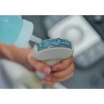
Zedu – what does it take to become a SEED?
What is a SEED? Where do SEEDs come from? How do SEEDs differ from a ‘traditional’ sonographer? What do you need to do to become one? Suean digs up some SEED information, which is emerging as a specialisation and career pathway in its own right.
 Medicine’s never been shy of a new acronym or abbreviation. AMI, IBS, ACL, SOB, RUQ; taken out of context, misunderstanding these can at best be humorous, at worst catastrophic. In recent years – at least since 2019 – yet another acronym has entered the Australian medical lexicon; the SEED.
Once (and might I say rather catchily) referred to as sonographer ‘proctored instruction in bedside ultrasound’[i], the role of SEEDs – or Sonographer Educators in the Emergency Department – has recently been formally recognised by the Australasian College of Emergency Medicine (ACEM) and its affiliate interest group Emergency Medicine Ultrasound Group (EMUGs).
[SEEDs are] experienced sonographers who bring technical knowledge, experience, and advanced psychomotor teaching skills into the Emergency Department to help with hands on training, reporting and assessments of Emergency Clinicians. Whilst their primary role will be in image acquisition and interpretation, there are increasing skills in clinical integration, such as with incidental or unexpected findings.[ii]
This definition serves two primary purposes.
- First it recognises the important and valuable technical skillset that a sonographer can bring to the emergency context and sits incontrovertibly within the traditional scope of practice – that of image acquisition and interpretation.
- Second it provides the basis for the creation of a SEED position description – foreshadowing key selection criteria that can be utilised by potential employers.
While the first purpose appeals to the ego (I mean who would question that sonographers are the recognised experts in image acquisition/interpretation), it’s the criteria within the second point that poses challenges – both to the emergency medicine and the sonographer communities alike.
As an ageing Holder of UltraSound Knowledge (HUSK) myself, the definition of a SEED is fundamentally aspirational, identifying the characteristics of what could be considered the ‘unicorn’ of sonographers. It’s the product of a world where – as access to ultrasound has increased – there has not been a concomitant increase in access to (or investment in) skilled and competent users of the technology in any specialty.
So as there has been a predictable increase in demands on sonographers – both in their traditional sphere and in expectations that they teach the art and science of imaging to other specialties – the SEED has emerged as a means to an end. And this situation isn’t unique to Australia, with counterparts in the USA also recognising the value that sonographers can deliver as a sonography education faculty member (SEFM).[iii]
Consequently, competition for sonographer time and skills has increased across the board; and so to have the opportunities.
But what does this mean for the sonographer chomping at the bit to leave the safety of radiology practice for the dynamism, challenges and reality of life in the world of emergency medicine? What does it take to make the transition to this role? Does it represent an easy option for sonographers to retire from the day-to-day grind? Or does it require a fundamentally different skillset that should rightly be recognised as a separate specialty? Let’s explore…
Unpicking the SEED
On many occasions I’ve been approached by sonographers tired of clinical life and looking for alternatives, either disaffected by the corporate grind or injured and facing a life changing decision to leave the profession.
 While the majority opt for a pre-retirement part time position, others consider teaching within the bounds of their ultrasound specialty – whether obstetrics, vascular or musculoskeletal. However, experience dictates that a sonographer tutor role is never simply that, and often devolves into running a room with (or without) a student. This ultimately leads to additional stress, injury and burnout. Add to this the increasing demands that covid-19 has put on every aspect of a sonographer’s daily life, and exit is an appealing option.
However – whether due to little exposure to POCUS, the negative connotations that remain around ultrasound users at the point of care or a lack of confidence – few consider a change in career of the likes that a SEED role offers.
Briefly summarising from the ACEM definition, a SEED candidate is one that:
- Is experienced
- Possesses technical knowledge, experience
- Holds advanced psychomotor teaching skills
- Will help with hands on training, reporting and assessments
- Is willing to increase skills in clinical integration, such as with incidental or unexpected findings
In the absence of a definitive position description, and with a few HUSKy years under my belt, here’s my take on unpicking the criteria and the challenges that emerge for anyone wanting to make the transition.
SEEDs and experience
Experience. A priceless aspect for any profession. It’s something earned – never bought. But what does this mean for a sonographer considering a SEED role? Does it simply mean the number of years you have been using ultrasound, or does it refer to something more esoteric such as being worldly wise?
 In my ‘experience’ hiring for attitude trumps formal qualifications every time. Attitude speaks to being open to new experiences, and the ability to draw on attributes and knowledge dynamically to meet a challenge – whether it be using a new type of ultrasound system each day or dealing with difficult patients. This will be fundamental for success as a SEED.
In the context of becoming a SEED, it’s also important to be mindful that while you may be experienced as a sonographer, this represents approximately 30 percent of the skill set necessary to succeed in a point-of-care teaching and training role, potentially less when we consider the learning curve necessary to get to grips with the specifics of emergency medicine and working in a dynamic team role.
We also must recognise that sonographers are trained to work independently and conditioned to report to a radiologist’s protocol, often specialising in a very narrow application. To transition from the role of ‘specialised documentary photojournalist’ to something more akin to a ‘focused sharpshooter’ where a yes/no answer immediately impacts patient management takes a shift in thinking that only comes with experience.
That’s why I’m of the view that a sonographer should have more than 5 years ‘time’ under their belt before they consider becoming a SEED. Time brings a confidence and maturity in troubleshooting that will be of immeasurable benefit in the role.
In the absence of a focused training pathway for SEEDs as a specialist role in its own right, it’ll take a combination of experience, the right attitude and a supportive team to make the change. This will most certainly be a challenge, but the rewards will be there.
SEEDs and technical knowledge
 To understand technical knowledge in ultrasound we should be clear that this can mean one of two things in the case of a SEED; it can either refer to competence in the specialist techniques (or art) of ultrasound in the context of emergency medicine, or the ability to utilise and optimise the tools (ultrasound technology and associated infrastructure) to practically solve problems.
From a sonographer’s – and even emergency physician – perspective this can easily be misconstrued as the easy bit in shifting from the radiology to point-of-care world. After all, surely from a technological perspective a linear probe’s a linear probe, and from a technical perspective abdomen scan an abdomen scan? However, it’s all too easy to underestimate the steepness of the learning curve associated with making the shift from radiology to point-of-care practitioner.
Technical knowledge – infrastructure
Let’s start with the comparatively simple notion of transferring your knowledge of ultrasound technology – probably better referred to as infrastructure knowledge. From an ultrasound system perspective physics remains an immutable fact; frequency, depth, focus and gain remain the same – so as a sonographer the fundamentals are well established. What may take some adjustment when moving from radiology to a SEED role is the type of system used, as well as how, where and when it’s used.
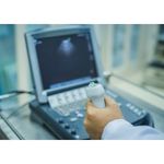 Designed for ease of use and robustness, point-of-care systems are the ultrasound equivalent of a point and press camera. While simple to use, don’t be fooled – their complexity should not be underestimated, and as technology advances their sophistication grows daily. You’ll need to become familiar with the wide variety of systems – their form factors, operating systems, strengths and weaknesses – and be comfortable jumping between handheld and more traditional cart style machines.
Your ultrasound knowledge will mean that as a SEED you’ll likely be involved in assisting with equipment purchasing decisions – ensuring that it will perform and do the job for which it’s intended. You’ll also potentially be responsible for liaising with system providers to ensure faults are rectified, maintenance scheduled and delivered. There may also be demands on you to assist with the curse of point-of-care ultrasound – that of image storage. Access to PACS is often a politically sensitive issue and may either result in you acting as negotiator for access, or establishing a stand-alone point-of-care image storage system. On all fronts, your technology skills will be tested.
Technical knowledge – technique
This, in my experience, is one of the biggest challenges when making the change from defining yourself as a sonographer to defining yourself as a SEED. While your ultrasound training provides you with the skills necessary to get and optimise an image, that same training can hinder your ability to be an effective SEED.
 From a technical perspective, sonographers are trained to take optimal images, shielded from the implications of diagnoses communicated by the radiologist to a referring clinician. In the point-of-care world, images are often not ideal and clinical decisions need to be made immediately. It is crucial to remember that a point-of-care exam is one that aims to answer a question with a binary answer – Yes or No. For example, is there free fluid in the abdomen – yes or no? Is there an intrauterine pregnancy – yes or no? If the clinical question requires more than binary answer, then it should be referred to the imaging department. Understanding the line between POCUS and formal imaging is crucial. Sometimes this is a matter of life and death; it’s raw and emotional work.
Consequently, as a SEED it’s imperative to consciously be utilitarian in approach, ditching technical perfectionism in favour of guiding physicians to be the best they can be. You’re there to help emergency physicians recognise when images are providing adequate information to assist in clinical management, when to call it quits, how to recognise their own limitations and not be afraid to point out errors. You’ll be constantly battling your inner perfectionist – you’ll need to get comfortable with your learners always leaving you feeling that their skills are not quite there. But remember, suboptimal images can be diagnostic. And given the relentless demands of emergency medicine, the reality is that those under your tutelage will never have the time to fully developing their imaging skills – and many often won’t think they need to. Your job as SEED will be to tread a fine line – ensuring your learners develop confidence and independence, while maintaining humility through self-reflective practice.
Equally, in order to maintain credibility, as a SEED you will need to learn from your learners and invest in expanding your clinical acumen (criteria five) in order to understand how visual information will be used – or not used – in patient management.
 You’ll need to become intimately familiar with and competent in the performance of the five examinations (and associated protocols/conventions) that are core to the emergency world: the eFAST (Extended Focused Assessment using Sonography in Trauma) exam, abdominal aortic aneurysm (AAA) assessment, basic echo in life support (BELS) imaging, lung assessment and procedural guidance (see Core Emergency Medicine Ultrasound Scans – a Sonographer’s Guide at the bottom of this post). While your ultrasound training will assist you in getting the views, you’ll need to expand your repertoire, work on your speed, and develop your confidence in order to transfer ultrasound skills to your learners.
Mastering each of these examinations and placing them in clinical context is hard work, part of an ongoing process that will level up your clinical knowledge and ultrasound skills as you develop as a SEED. The learning curve as you develop a methodology to teach and support your learners will be steep and at times frustrating but the rewards – in seeing your skills and those of your charges develop – immeasurable.
SEEDs and advanced psychomotor teaching skills, hands on training, reporting and assessments
 Combining criteria three and four, the most significant academic challenge facing any SEED will certainly be that of curriculum development, working to create a program of sequential learning tasks that result in physicians developing as independent and safe users of ultrasound.
In most instances, this will be a completely foreign concept to sonographers. As an advanced teacher, a SEED will be expected to identify learning objectives, create lesson plans, conduct skills audits, develop formal assessment and feedback processes, evolve quality assurance programs, design and deliver presentations – all but a snapshot of what will be required to ensure the oversight and delivery of an appropriately structured formal ultrasound program.
What I find particularly fascinating is the specific focus on holding advanced psychomotor teaching skills, which is still an emerging concept in the teaching of ultrasound.
Psychomotor skills in the context of ultrasound imaging was first defined in ground breaking research by Nicholls et. al. as ‘the unique mental and motor activities required to execute a manual task safely and efficiently for each clinical situation’.[iv] In Nicholls’ paper, psychomotor skills (open and closed) are of two types – visuomotor and visuospatial – whose mastery by an ultrasound student are considered fundamental to achieving competency.
As a theory still in its emergent phase, the potential implications for ultrasound teaching – making conscious the intuitive and subconscious skills held by sonographers and determining how to transfer that knowledge to new learners – have yet to be fully explored.
As the concept of psychomotor skills (and its implications on learning ultrasound) is not taught to sonographers, it will be incumbent on any SEED to become familiar with the theory and how its concepts can be used to derive and refine a teaching program, all the while ensuring clinical relevance and skills development for the learner. The SEED that cracks this code will truly demarcate the difference between a sonographer overseer and a sonographer educator.
Other observations for SEEDs
My final observations of what it means to be a SEED turn to the many challenges and opportunities posed by cultural change.
 Leaving the secure orbit of a radiologist to pursue a career in emergency medicine will be equal parts scary and thrilling. Instead of working alone in a dark room, you’ll be part of a dynamic team where your opinion is valued and relied upon, but conversely you will be held responsible for your decisions.
As a SEED, you can’t afford to be intimidated by the cultural conventions of ‘hierarchy’, where physicians are considered higher in the ‘pecking order’ than sonographers. Remember – you are the imaging expert – so it’s inevitable that you will need to correct a doctor’s technique, or even deem that they are not yet competent to perform a certain exam. However, as much as you can’t be intimidated, it’s also important to remember that you are not a doctor and that in clinical decision-making ultrasound is just a small part of a larger equation. It’s a fine line to tread to ensure that POCUS is performed safely and responsibly.
Finally, as someone who has been teaching POCUS for over 15 years, be prepared to face some opposition and even hostility from other clinical sonographers. Not everyone is on board with the concept that point-of-care ultrasound legitimately deserves a place at the table. Those that aren’t often don’t truly understand what the goals of POCUS are or how it can so positively impact the patient journey. Things are changing ever so slowly but the resistance – whether driven by politics, lack of knowledge or even disinformation – still rumbles.
But never lose sight of the potential. If you can survive the culture shock and weather the politics, you’ll get the opportunity to spread your wings as a new career path reveals itself.
SEED – it’s a whole new world
Reflecting on my experience as an educator in the emergency space, and the developments that we have seen in recent times, pursuing a career as a SEED has the potential to be an amazingly rewarding experience that will challenge you immensely.
However, it must be said that being a SEED is not right for everyone. In the current environment it’s tempting for a sonographer to regard the position as an ‘easy out’ of clinical practice, or a semi-retirement options for those with injuries. However, this couldn’t be further from the truth. Given the need to learn new technologies, new techniques and clinical applications, develop academically sound education programs and grow your managerial skills – in many ways becoming a SEED and the unique combination of skills and abilities that you will need to develop means it should in fact be recognised as a distinct career pathway.
If that’s you, may your seed bloom.
Core Emergency Medicine Ultrasound Scans – a Sonographer’s Guide
As a SEED you’ll need to become intimately familiar with and teach each of these core emergency ultrasound scans – their purpose and implications for patient management, the pitfalls and limitations of each, the tips and tricks that will allow physicians to make a diagnostic call – all the while recognising the limitations of that the operating environment and the unique presentation of patients in an emergent context.
eFAST – Extended Focussed Abdominal Sonography for TraumaThe eFAST exam is a protocol comprised of six views used to detect the presence of free fluid /blood in the abdomen or thorax in patients who’ve been subject to blunt trauma. Some views are also used to detect free fluid in the case of ruptured ectopic pregnancy. The views include the right upper quadrant (RUQ), left upper quadrant (LUQ), pelvis and the subcostal four chamber view of the heart. The final views are of the anterior chest wall to exclude pneumothorax and the base of the lungs to exclude haemothorax. |
AAA – Abdominal Aortic Aneurysm assessmentSonographers are familiar with the conventional assessment for an abdominal aneurysm, however the exam is truncated in the emergency context (patients don’t come prepped for the exam!) Doctors in a hurry make different mistakes. |
BELS – The Basic Echo in Life Support examinationThe BELS exam includes four basic views of the heart – parasternal long axis (PLAX), parasternal short axis (PSAX), apical four chamber (AP4) and subcostal four chamber (SC4) – performed with the aim of detecting signs that the heart is still beating (or pulseless electrical activity), and/or pericardial effusion and tamponade. Additional but non-imperative information includes identifying major wall motion defects, left ventricular function, right ventricular function, and signs of pulmonary embolism. This examination is predominantly a B-mode assessment of the heart performed in under 5 minutes which provides enough information to rule out major conditions and to guide treatment. Very few emergency physicians have advanced cardiac imaging ability or perform the numbers required to be able to use Doppler well and quickly enough in their assessment of the heart. |
LungLung ultrasound – once considered impossible by sonographers – is super easy to perform but much harder to interpret. It requires a good understanding of the pathological conditions that affect the lung and how those pathologies interact with sound. Unlike other ultrasound imaging – where we rely on interpreting how the sound reflects in different ways from the various tissue – in lung ultrasound it’s all about understanding the implications of the signature artifacts and what that means clinically. There are many different protocols, determined by the suspected pathology, the condition of the patient and patient positioning. Lung ultrasound protocols contain a lot of confusing terminology, with many different terms used to describe the same thing. |
Procedural GuidanceThe key to teaching procedural guidance is to have a solid understanding of each procedure ultrasound guided needle placement will be used for, and to understand the terminology. Procedures include central and peripheral venous access, nerve blocks, pleural taps, lumbar punctures and more. The principle for guiding the needle is the same wherever the needle ends up, however potential complications differ for each procedure. Understanding complications guides selection of needle approach – either in-plane or out of plane. From an ultrasound perspective, it’s a simple process, but foreign to the clinical sonographer because this is usually the radiologist’s domain. |
Endnotes
[i] Sweetman G, Fear M, Hird K. Experience of a tutor centric model for sonography training of emergency department registrars in an Australian urban emergency department 2009-2012. Australasian Journal of Ultrasound in Medicine. 2015;18(3):112-117.
[ii] Australasian College for Emergency Medicine (ACEM) & Emergency Medicine Ultrasound Group(EMUGs). Sonographer Educators in Emergency Departments (SEEDS) Definition. V1 December 2021.
[iii] Grimmett S, End B, Quedado K, Kraft C, Minardi J. The Value of Sonography Education Faculty in an Academic Emergency Ultrasound Program. Journal of Diagnostic Medical Sonography. 2020;36(5): 507–510.
[iv] Nicholls D, Sweet L, Hyett J. Psychomotor skills in medical ultrasound imaging: an analysis of the core skill set. J Ultrasound Med. 2014 Aug;33(8):1349-52. doi: 10.7863/ultra.33.8.1349. PMID: 25063399.
You may also like

Zedu Weekly Wrap – 26 April 2024

Zedu Weekly Wrap – 19 April 2024
